. 1998 Apr;180(7):1904-12.
doi: 10.1128/JB.180.7.1904-1912.1998.

An export-specific reporter designed for gram-positive bacteria: application to Lactococcus lactis

Affiliations

An export-specific reporter designed for gram-positive bacteria: application to Lactococcus lactis

I Poquet et al. J Bacteriol. 1998 Apr.

Erratum in

  • J Bacteriol 1998 Aug;180(16):4324

Abstract

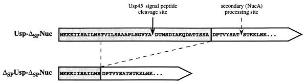
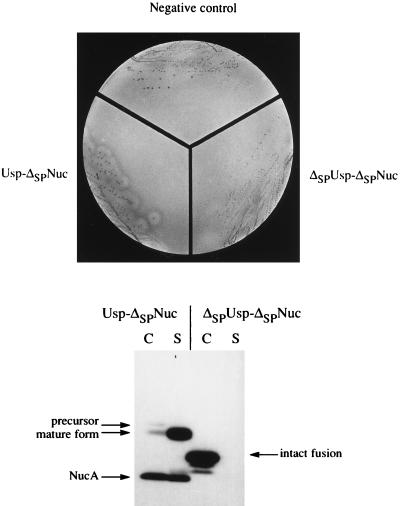
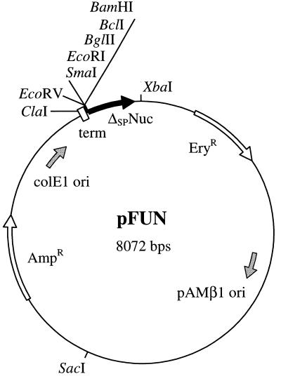
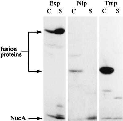
The identification of exported proteins by fusion studies, while well developed for gram-negative bacteria, is limited for gram-positive bacteria, in part due to drawbacks of available export reporters. In this work, we demonstrate the export specificity and use of the Staphylococcus aureus secreted nuclease (Nuc) as a reporter for gram-positive bacteria. Nuc devoid of its export signal (called delta(SP)Nuc) was used to create two fusions whose locations could be differentiated. Nuclease activity was shown to require an extracellular location in Lactococcus lactis, thus demonstrating the suitability of delta(SP)Nuc to report protein export. The shuttle vector pFUN was designed to construct delta(SP)Nuc translational fusions whose expression signals are provided by inserted DNA. The capacity of delta(SP)Nuc to reveal and identify exported proteins was tested by generating an L. lactis genomic library in pFUN and by screening for Nuc activity directly in L. lactis. All delta(SP)Nuc fusions displaying a strong Nuc+ phenotype contained a classical or a lipoprotein-type signal peptide or single or multiple transmembrane stretches. The function of some of the predicted signals was confirmed by cell fractionation studies. The fusions analyzed included long (up to 455-amino-acid) segments of the exported proteins, all previously unknown in L. lactis. Homology searches indicate that several of them may be implicated in different cell surface functions, such as nutrient uptake, peptidoglycan assembly, environmental sensing, and protein folding. Our results with L. lactis show that delta(SP)Nuc is well suited to report both protein export and membrane protein topology.


Figures

FIG. 1
FIG. 1
N-terminal sequences of Usp-ΔSPNuc and ΔSPUsp-ΔSPNuc fusion proteins. Sequences derived from Usp45 or from ΔSPNuc (i.e., C-terminal 155 amino acids of Nuc) are shown in shaded or white boxes, respectively. The solid arrow indicates the Usp45 signal peptide cleavage site (61). The dashed arrow indicates the secondary site of proteolysis, which results in the NucA form (i.e., C-terminal 147 amino acids of Nuc) (30). The dashed lines joining Usp-ΔSPNuc and ΔSPUsp-ΔSPNuc indicate the region of the former fusion which is absent in the latter.
FIG. 2
FIG. 2
Specific detection of ΔSPNuc fusions which have an export signal in L. lactis. (Top) Nuc activity of Usp-ΔSPNuc but not of ΔSPUsp-ΔSPNuc is detected by plate tests. MG1363 strains containing plasmid pFUN (negative control), pVE8009 (Usp-ΔSPNuc), or pVE8010 (ΔSPUsp-ΔSPNuc) were streaked onto solid medium (brain heart infusion agar) and grown overnight. Colonies were overlaid with indicator medium containing toluidine blue, denatured DNA, and agar. Nuclease activity is detected by pink halos around colonies (Nuc+ phenotype) only in the case of MG1363(pVE8009), which produces Usp-ΔSPNuc. (Bottom) Location of Usp-ΔSPNuc and ΔSPUsp-ΔSPNuc fusion proteins in L. lactis. Cell (C) and supernatant (S) fractions of mid-exponential-phase cultures of L. lactis MG1363 derivative strains producing either Usp-ΔSPNuc or ΔSPUsp-ΔSPNuc fusion protein (from plasmids pVE8009 and pVE8010, respectively) were analyzed by SDS-PAGE and Western blotting using polyclonal Nuc antibodies. Putative forms of each fusion are indicated. Commercial NucA, used as a size reference (not presented), comigrates with the smallest degradation product of Usp-ΔSPNuc. Note that aberrant relative migration of Usp-ΔSPNuc forms (precursor, mature, and NucA) has previously been observed (30, 31) and that NucA in the cell fraction may be cytoplasmic or externally cell associated (31).
FIG. 3
FIG. 3
pFUN, a new probe vector for identification of genes encoding exported proteins. pFUN is an 8,072-bp shuttle vector that contains pAMβ1 (active in various lactic acid bacteria) and ColE1 (active in E. coli) replicons. A multicloning site (BamHI, BclI, BglII, EcoRI, and SmaI) allows the creation of translational fusions between genomic DNA fragments and the ORF (Δnuc) for ΔSPNuc (black arrow). If the inserted DNA supplies transcriptional and translational signals, a fusion with ΔSPNuc could be produced, and if the polypeptide fused to ΔSPNuc supplies an export signal, the fusion would display Nuc activity. Note that BamHI, BclI, and BglII sites were designed to allow fusions in the three ORFs and that a unique EcoRV site upstream of the multicloning site is also available. The trpA ρ-independent terminator (term) ensures that transcription of the translational fusion initiates from signals present on the inserted DNA. Unique restriction sites, genes conferring antibiotic resistance (open arrows on the plasmid), and origins of replication (grey arrows inside the plasmid) are shown. Further details of construction are given in Materials and Methods. AmpR, ampicillin resistance marker; EryR, erythromycin resistance marker.
FIG. 4
FIG. 4
Location of Exp1-ΔSPNuc, Nlp1-ΔSPNuc, and Tmp2-ΔSPNuc fusion proteins in L. lactis. Three L. lactis MG1363 derivative strains, each producing a representative member of each class of putative exported ΔSPNuc fusions (shown in parentheses), were examined: Exp (Exp1-ΔSPNuc), Nlp (Nlp1-ΔSPNuc), and Tmp (Tmp2-ΔSPNuc). Cell (C) and supernatant (S) fractions from mid-exponential-phase cultures of the above-mentioned strains were analyzed by SDS-PAGE and Western blotting using polyclonal Nuc antibodies. Putative forms of each fusion are indicated. Note that a band corresponding to a high-molecular-weight protein, visible in the cell fraction of the strain producing Nlp1-ΔSPNuc, is nonspecific and is observed in other samples.

References

    1. Ahrenholtz I, Lorenz M G, Wackernagel W. A conditional suicide system in Escherichia coli based on the intracellular degradation of DNA. Appl Environ Microbiol. 1994;60:3746–3751. - PMC - PubMed
    1. Baba T, Schneewind O. Target cell specificity of a bacteriocin molecule: a C-terminal signal directs lysostaphin to the cell wall of Staphylococcus aureus. EMBO J. 1996;15:4789–4797. - PMC - PubMed
    1. Bardwell J C, McGovern K, Beckwith J. Identification of a protein required for disulfide bond formation in vivo. Cell. 1991;67:581–589. - PubMed
    1. Bolhuis H, Poelarends G, van Veen H W, Poolman B, Driessen A J, Konings W N. The lactococcal lmrP gene encodes a proton motive force-dependent drug transporter. J Biol Chem. 1995;270:26092–26098. - PubMed
    1. Broome-Smith J K, Tadayyon M, Zhang Y. β-Lactamase as a probe of membrane protein assembly and protein export. Mol Microbiol. 1990;4:1637–1644. - PubMed

MeSH terms

LinkOut - more resources