. 1998 Apr 14;95(8):4140-5.
doi: 10.1073/pnas.95.8.4140.

The structure of the two amino-terminal domains of human ICAM-1 suggests how it functions as a rhinovirus receptor and as an LFA-1 integrin ligand

Affiliations

The structure of the two amino-terminal domains of human ICAM-1 suggests how it functions as a rhinovirus receptor and as an LFA-1 integrin ligand

J Bella et al. Proc Natl Acad Sci U S A. .

Abstract

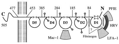
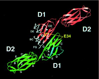
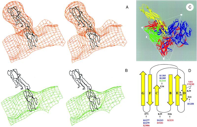
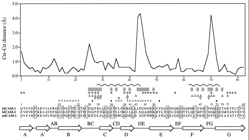
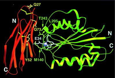
The normal function of human intercellular adhesion molecule-1 (ICAM-1) is to provide adhesion between endothelial cells and leukocytes after injury or stress. ICAM-1 binds to leukocyte function-associated antigen (LFA-1) or macrophage-1 antigen (Mac-1). However, ICAM-1 is also used as a receptor by the major group of human rhinoviruses and is a catalyst for the subsequent viral uncoating during cell entry. The three-dimensional atomic structure of the two amino-terminal domains (D1 and D2) of ICAM-1 has been determined to 2.2-A resolution and fitted into a cryoelectron microscopy reconstruction of a rhinovirus-ICAM-1 complex. Rhinovirus attachment is confined to the BC, CD, DE, and FG loops of the amino-terminal Ig-like domain (D1) at the end distal to the cellular membrane. The loops are considerably different in structure to those of human ICAM-2 or murine ICAM-1, which do not bind rhinoviruses. There are extensive charge interactions between ICAM-1 and human rhinoviruses, which are mostly conserved in both major and minor receptor groups of rhinoviruses. The interaction of ICAMs with LFA-1 is known to be mediated by a divalent cation bound to the insertion (I)-domain on the alpha chain of LFA-1 and the carboxyl group of a conserved glutamic acid residue on ICAMs. Domain D1 has been docked with the known structure of the I-domain. The resultant model is consistent with mutational data and provides a structural framework for the adhesion between these molecules.


Figures

Figure 1
Figure 1
A diagram of an ICAM-1 molecule showing sites of glycosylation (lollipop-shaped structures) and the approximate location of binding sites of LFA-1, Mac-1, human rhinoviruses, fibrinogen, and Plasmodium falciparum-infected erythrocytes (PFIE).
Figure 2
Figure 2
The crystallographic dimer in which there is extensive antiparallel β-sheet between the G strands. The residues and loops that penetrate into the HRV “canyon” are colored white in one of the two monomers. Glu-34, essential for LFA-1 binding, is shown in yellow. Strands and loops important for binding PFIE are in light blue in one monomer. β-strands in domains D1 and D2 are labeled consistent with the nomenclature of an “Intermediate” and “Constant 2” Ig-like fold, respectively (25).
Figure 3
Figure 3
Interpretation of the cryo-EM electron density for HRV16 complexed with a D1D2 fragment of ICAM-1 expressed in Chinese hamster ovary cells (14). (A) Stereoview of the cryo-EM electron density of the complex (orange) fitted with the mutICAM-1 Cα backbone. The extra electron density regions around D2 of ICAM-1 correspond to the predicted locations of the four glycosylation sites. Domain D1 is not glycosylated. (B) Stereoview of the cryo-EM density of HRV16 (green) with the ICAM-1 Cα backbone, which can be seen to fit into the canyon depression on the HRV16 surface. (C) Ribbon diagram showing the interaction of ICAM-1 (yellow) and HRV16. HRV16 proteins VP1, VP2, and VP3 are in blue, green, and red, respectively. Two symmetry-related VP1s and VP3s are shown. Some icosahedral symmetry elements and the boundary of an icosahedral asymmetric unit are shown. (D) Diagrammatic figure of the fold of ICAM-1 domain D1 (yellow). ICAM-1 residues that could make salt bridges in the HRV16 complex are in black. The opposing residues on HRV16 are colored blue, green, and red and are numbered starting at 1001, 2001, 3001 according to whether they are in viral proteins VP1, VP2, or VP3, respectively. There are 60 copies of each viral protein in the HRV16 complex.
Figure 4
Figure 4
Structural alignment of D1 of mutICAM-1 with that of ICAM-2. The plot is of differences in Cα positions for the superimposed structures. By far, the largest differences occur at the BC, DE, and FG loops, all of which are at the amino end of the domain and important for binding of HRVs to human (h) ICAM-1 [but not human (h) ICAM-2 or murine (m) ICAM-1]. Residues below the gray bars interact with HRV16 when mutICAM-1 is fitted into the cryo-EM electron density map of the virus–receptor complex. ICAM-1 residues identified by mutational studies as being involved in binding HRVs are marked with ∗ (11), ▵ (12), and ▿ (13). Residues marked with ♦ have been identified by single amino acid mutations as being important in binding LFA-1 (24). Residues probably involved in binding to PFIE are marked with × (11). The wavy line (∼) indicates residues that interact with LFA-1 for the docking shown in Fig. 5. Sequence identities are shown in boxes.
Figure 5
Figure 5
Ribbon diagram showing docking of the I-domain of LFA-1 (green) with domain D1 of mutICAM-1 (orange). Coordination of the metal ion (purple) on the I-domain is completed by Glu-34 (white) on the β-strand C of mutICAM-1. Additional residues of the I-domain (36) and of ICAM-1 (24) considered important for binding are shown in green and yellow, respectively.

Similar articles

Cited by

References

    1. van de Stolpe A, van der Saag P T. J Mol Med. 1996;74:13–33. - PubMed
    1. Greve J M, Davis G, Meyer A M, Forte C P, Yost S C, Marlor C W, Kamarck M E, McClelland A. Cell. 1989;56:839–847. - PubMed
    1. Staunton D E, Merluzzi V J, Rothlein R, Barton R, Marlin S D, Springer T A. Cell. 1989;56:849–853. - PubMed
    1. Berendt A R, McDowall A, Craig A G, Bates P A, Sternberg M J E, Marsh K, Newbold C I, Hogg N. Cell. 1992;68:71–81. - PubMed
    1. Ockenhouse C F, Betageri R, Springer T A, Staunton D E. Cell. 1992;68:63–69. - PubMed

Publication types

MeSH terms