

Telomere loss in cells treated with cisplatin
- PMID: 9539717
- PMCID: PMC22469
- DOI: 10.1073/pnas.95.8.4219
Telomere loss in cells treated with cisplatin
Abstract
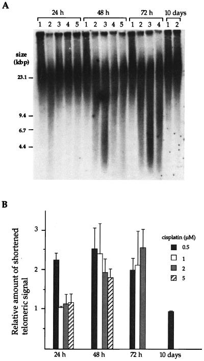
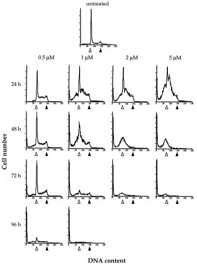
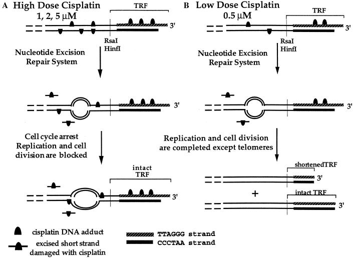
Telomeres play an important role in the immortalization of proliferating cells. The long tandem repeats of 5'-TTAGGG-3' sequences in human telomeres are potential targets for the anticancer drug cisplatin, which forms mainly intrastrand d(GpG) and d(ApG) cross-links on DNA. The present study reveals that telomeres in cisplatin-treated HeLa cells are markedly shortened and degraded. A dose that killed 61% of the cells but allowed one round of cell division resulted in shortened telomeres before the induction of apoptosis. Higher doses of cisplatin halted cell cycle progression during the first S phase and triggered apoptosis followed by degradation of telomere repeats. A model in which both cell division with incomplete replication and induction of apoptosis by cisplatin could occur was devised to explain the drug-induced telomere loss.
Figures

Similar articles
-
Tumour-cell apoptosis after cisplatin treatment is not telomere dependent.Int J Cancer. 2006 Jun 1;118(11):2727-34. doi: 10.1002/ijc.21675. Int J Cancer. 2006. PMID: 16381006
-
Formation of cis-diamminedichloroplatinum(II) 1,2-intrastrand cross-links on DNA is flanking-sequence independent.Nucleic Acids Res. 2000 Nov 1;28(21):4237-43. doi: 10.1093/nar/28.21.4237. Nucleic Acids Res. 2000. PMID: 11058123 Free PMC article.
-
The interaction of cisplatin with a human telomeric DNA sequence containing seventeen tandem repeats.Bioorg Med Chem Lett. 2013 Feb 15;23(4):1041-5. doi: 10.1016/j.bmcl.2012.12.021. Epub 2012 Dec 20. Bioorg Med Chem Lett. 2013. PMID: 23302441
-
[Regulation of telomeres length: making the telomeres accessible?].Bull Cancer. 2005 Jan;92(1):13-22. Bull Cancer. 2005. PMID: 15811846 Review. French.
-
[Linear prokaryotic chromosome; structure and synthesis of telomeres].Postepy Biochem. 1997;43(1):12-7. Postepy Biochem. 1997. PMID: 9289722 Review. Polish. No abstract available.
Cited by
-
Sodium butyrate enhances the cytotoxic effect of cisplatin by abrogating the cisplatin imposed cell cycle arrest.BMC Mol Biol. 2010 Jun 24;11:49. doi: 10.1186/1471-2199-11-49. BMC Mol Biol. 2010. PMID: 20576112 Free PMC article.
-
Dual functional dinuclear platinum complex with selective reactivity towards c-myc G-quadruplex.Sci Rep. 2018 Jan 15;8(1):767. doi: 10.1038/s41598-017-19095-y. Sci Rep. 2018. PMID: 29335501 Free PMC article.
-
Molecular disruption of RAD50 sensitizes human tumor cells to cisplatin-based chemotherapy.J Clin Invest. 2009 Jul;119(7):1974-85. doi: 10.1172/JCI33816. J Clin Invest. 2009. PMID: 19487811 Free PMC article.
-
DNA interstrand cross-link repair requires replication-fork convergence.Nat Struct Mol Biol. 2015 Mar;22(3):242-7. doi: 10.1038/nsmb.2956. Epub 2015 Feb 2. Nat Struct Mol Biol. 2015. PMID: 25643322 Free PMC article.
-
Platination of telomeric DNA by cisplatin disrupts recognition by TRF2 and TRF1.J Biol Inorg Chem. 2010 Jun;15(5):641-54. doi: 10.1007/s00775-010-0631-4. Epub 2010 Feb 27. J Biol Inorg Chem. 2010. PMID: 20191372
References
-
- Pil P, Lippard S J. Cisplatin and Related Drugs. San Diego: Academic; 1997. pp. 392–410.
-
- Chu G. J Biol Chem. 1994;269:787–790. - PubMed
-
- Lepre C A, Lippard S J. Nucleic Acids Mol Biol. 1990;4:9–38.
-
- Zamble D B, Mu D, Reardon J T, Sancar A, Lippard S J. Biochemistry. 1996;35:10004–10013. - PubMed
-
- Fraval H N A, Rawlings C J, Roberts J J. Mutat Res. 1978;51:121–132. - PubMed
Publication types
MeSH terms
Substances
Grants and funding
LinkOut - more resources
Full Text Sources
Other Literature Sources
