. 1998 Apr 14;95(8):4224-8.
doi: 10.1073/pnas.95.8.4224.

Amyloid fibril formation by an SH3 domain

Affiliations

Amyloid fibril formation by an SH3 domain

J I Guijarro et al. Proc Natl Acad Sci U S A. .

Abstract

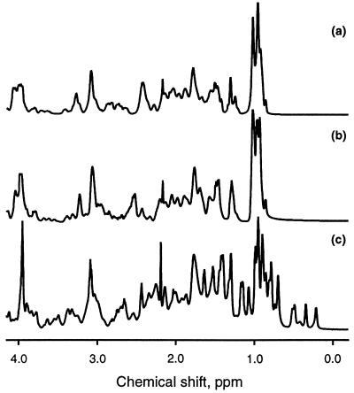
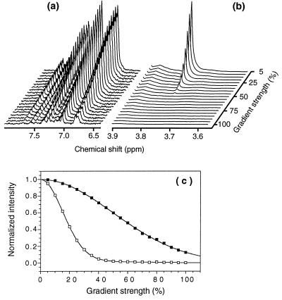
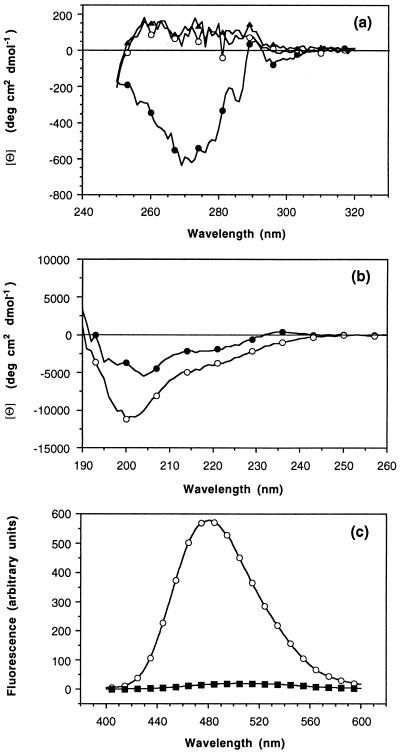
The SH3 domain is a well characterized small protein module with a simple fold found in many proteins. At acid pH, the SH3 domain (PI3-SH3) of the p85alpha subunit of bovine phosphatidylinositol 3-kinase slowly forms a gel that consists of typical amyloid fibrils as assessed by electron microscopy, a Congo red binding assay, and x-ray fiber diffraction. The soluble form of PI3-SH3 at acid pH (the A state by a variety of techniques) from which fibrils are generated has been characterized. Circular dichroism in the far- and near-UV regions and 1H NMR indicate that the A state is substantially unfolded relative to the native protein at neutral pH. NMR diffusion measurements indicate, however, that the effective hydrodynamic radius of the A state is only 23% higher than that of the native protein and is 20% lower than that of the protein denatured in 3.5 M guanidinium chloride. In addition, the A state binds the hydrophobic dye 1-anilinonaphthalene-8-sulfonic acid, which suggests that SH3 in this state has a partially formed hydrophobic core. These results indicate that the A state is partially folded and support the hypothesis that partially folded states formed in solution are precursors of amyloid deposition. Moreover, that this domain aggregates into amyloid fibrils suggests that the potential for amyloid deposition may be a common property of proteins, and not only of a few proteins associated with disease.


Figures

Figure 1
Figure 1
Electron micrograph of a negatively stained preparation of PI3-SH3 fibrils at pH 2.0. (Bar = 1,000 Å.)
Figure 2
Figure 2
X-ray diffraction pattern of PI3-SH3 fibrils. The reflections generated by the interstrand spacing (4.7 Å) and the intersheet spacing (centered at 9.4 Å) typical of amyloid fibrils are marked by arrows. Because of poor alignment of the fibrils, these reflections appear as rings and do not display a meridional or equatorial character.
Figure 3
Figure 3
One-dimensional NMR spectra of the denatured protein (D) in 3.5 M GuHCl, pH 7.2 (a), of the acid state (A) at pH 2.0 (b), and of the native protein (N) at pH 7.2 (c).
Figure 4
Figure 4
Aromatic region (a) and expansion of the dioxane signal (b) of the 1H NMR spectra of the PI3-SH3 A state obtained with the PG-SLED sequence at different pulse field-gradient strengths. The signal of the small reference molecule dioxane decays much faster than the signals from the protein. (c) Dependence of the integral of the aromatic region between 6.6 and 7.7 ppm (▪) and of the dioxane (□) signal (3.75 ppm) on the strength of the field gradient, expressed as a percentage of the maximum strength used (≈60 G cm−1). Data shown are derived from experiments performed on a PI3-SH3 sample (0.3 mM) at pH* 2.0 and 20°C. Analogous curves were obtained for the native protein and for the protein denatured in 3.5 M GuHCl. The solid lines in c correspond to fits to a Gaussian function (for the protein aromatic signals) or to a sum of two Gaussians (for the dioxane signal that resonates at a frequency where signals arising from the protein also resonate) (see ref. 26). Fitted decay rates are proportional to the diffusion coefficients for the protein and dioxane molecules, which are inversely proportional to their effective hydrodynamic radii.
Figure 5
Figure 5
CD spectra of PI3-SH3 in the near- (a) and far-UV (b) regions (20°C). Native state, pH 7.2 (•); A state, pH 2.0 (○); and denatured state in 3.5 M GuHCl (▴). For the near-UV spectra, the ellipticity is expressed in terms of molar ellipticity per aromatic residue. Protein concentration was determined spectrophotometrically by using an extinction coefficient of 1.896 (mg/ml)−1 cm−1 for the native and A states and of 2.011 (mg/ml)−1 cm−1 for the denatured protein. Extinction coefficients were determined as described in ref. . (c) Fluorescence emission spectra of ANS (250 μM) in the presence of 15 μM PI3-SH3 (○) and without protein (▪) acquired on pH 2.0 samples at 20°C. The excitation wavelength was 370 nm, and the excitation and emission monochromator slit widths were both set to 5 nm.

Similar articles

Cited by

References

    1. Tan S Y, Pepys M B. Histopathology. 1994;25:403–414. - PubMed
    1. Kelly J W. Curr Op Struct Biol. 1996;6:11–17. - PubMed
    1. Sunde M, Blake C C F. Adv Protein Chem. 1997;50:123–159. - PubMed
    1. Blake C, Serpell L C. Structure. 1996;4:989–998. - PubMed
    1. Sunde M, Serpell L C, Bartlam M, Fraser P E, Pepys M B, Blake C C F. J Mol Biol. 1997;273:729–739. - PubMed

Publication types

LinkOut - more resources