. 1998 Apr 14;95(8):4309-14.
doi: 10.1073/pnas.95.8.4309.

NMR determination of the major solution conformation of a peptoid pentamer with chiral side chains

Affiliations

NMR determination of the major solution conformation of a peptoid pentamer with chiral side chains

P Armand et al. Proc Natl Acad Sci U S A. .

Abstract

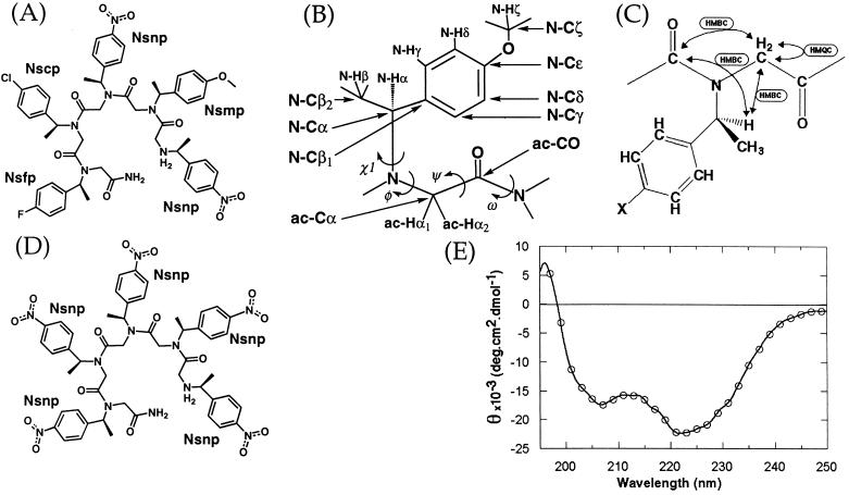
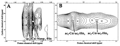
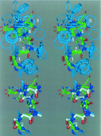
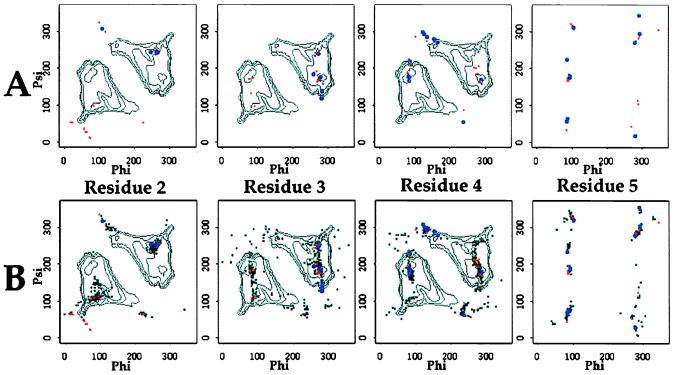
Polymers of N-substituted glycines ("peptoids") containing chiral centers at the alpha position of their side chains can form stable structures in solution. We studied a prototypical peptoid, consisting of five para-substituted (S)-N-(1-phenylethyl)glycine residues, by NMR spectroscopy. Multiple configurational isomers were observed, but because of extensive signal overlap, only the major isomer containing all cis-amide bonds was examined in detail. The NMR data for this molecule, in conjunction with previous CD spectroscopic results, indicate that the major species in methanol is a right-handed helix with cis-amide bonds. The periodicity of the helix is three residues per turn, with a pitch of approximately 6 A. This conformation is similar to that anticipated by computational studies of a chiral peptoid octamer. The helical repeat orients the amide bond chromophores in a manner consistent with the intensity of the CD signal exhibited by this molecule. Many other chiral polypeptoids have similar CD spectra, suggesting that a whole family of peptoids containing chiral side chains is capable of adopting this secondary structure motif. Taken together, our experimental and theoretical studies of the structural properties of chiral peptoids lay the groundwork for the rational design of more complex polypeptoid molecules, with a variety of applications, ranging from nanostructures to nonviral gene delivery systems.


Figures

Figure 1
Figure 1
(A) Compound 1 (Nsnp1-Nsmp2-Nsnp3-Nscp4-Nsfp5). Nsnp = (S)-N-(1-(p-nitrophenylethyl))glycine, Nsmp = (S)-N-(1-(p-methoxyphenylethyl))glycine, Nscp = (S)-N-(1-(p-chlorophenylethyl))glycine, Nsfp = (S)-N-(1-(p-fluoro-phenylethyl))glycine. The amide bonds are drawn in their cis configurations. (B) Atom and dihedral nomenclature. These are shown for Nsmp. The nomenclatures for the other residues are identical except for the absence of Nsmp-Cζ and Nsmp-Hζ atoms. Atom names follow (15); however, the side chain name subscript has been dropped when not referring to a particular residue (e.g., N-Cα instead of Nxxx-Cα) and has been omitted in this figure for clarity. Dihedral angles are named by analogy to those of peptides. (C) Simplified schematic representation of the assignment process for part of 1. Arrows indicate the connections that were used and the spectrum types with which they were established. (D) Pentamer of (S)-N-(1-(p-nitrophenylethyl))glycine [(Nsnp)5]. (E) CD spectrum of 1 in 100% methanol. The data were collected at 10°C and at a concentration of 0.1 mM.
Figure 2
Figure 2
Detail of the heteronuclear spectra used to assign the resonances of the main chain and side chain α protons of residue 1. The spectra were collected on a molecule containing 13C labels at the ac1-Cα and ac1-CO positions. (A) HMBC spectrum showing the ac1-Cα-Nsnp1-Hα cross peak for the major isomer. (B) HMQC spectrum showing the ac1-Cα-Nsnp1-Hα cross peaks for the major isomer.
Figure 3
Figure 3
Stereo representations of the conformations remaining after restraint violation analysis. (Upper) Cluster A. This describes the major conformation of 1 in methanol. Backbone carbons are shown in green, side chain carbons in light blue, nitrogen atoms in dark blue, and oxygen atoms in red. The para substituents on the aromatic rings and the hydrogen atoms have been omitted for clarity. Residue numbers are shown in white. (Lower) Comparison of cluster A conformations with previously published model (6). Only backbone atoms and side chain α carbons are shown. The model is shown as a thicker ball and stick molecule, and its carbon atoms are colored white.
Figure 4
Figure 4
Distribution of the values of the main chain dihedral angles (φ and ψ) for residues 2 to 5 (residue 1 is not included because there is no proper φ dihedral angle). (A) Cluster A (large blue circles) and clusters B–H (small red circles) after minimization and before ensemble calculation. (B) Same, with cluster A angles after ensemble calculation (small black circles). White contour lines for residues 2–4 indicate the potential surface for Nsnp oligomers, after ref. .

Similar articles

Cited by

References

    1. Simon R J, Kania R S, Zuckermann R N, Huebner V D, Jewell D A, et al. Proc Natl Acad Sci USA. 1992;89:9367–9371. - PMC - PubMed
    1. Zuckermann R N, Martin E J, Spellmeyer D C, Stauber G B, Shoemaker K R, et al. J Med Chem. 1994;37:2678–2685. - PubMed
    1. Zuckermann R N, Kerr J M, Kent S B H, Moos W H. J Am Chem Soc. 1992;114:10646–10647.
    1. Murphy J E, Uno T, Hamer J D, Cohen F E, Dwarki V, Zuckermann R N. Proc Natl Acad, Sci USA. 1997;95:1517–1522. - PMC - PubMed
    1. Kirshenbaum K, Barron A E, Goldsmith R E, Armand P A, Bradley E K, Truong K T V, Dill K A, Cohen F E, Zuckermann R N. Proc Natl Acad Sci USA. 1997;95:4303–4308. - PMC - PubMed

Publication types

LinkOut - more resources