

Evidence for keratinocyte stem cells in vitro: long term engraftment and persistence of transgene expression from retrovirus-transduced keratinocytes
- PMID: 9539741
- PMCID: PMC22493
- DOI: 10.1073/pnas.95.8.4356
Evidence for keratinocyte stem cells in vitro: long term engraftment and persistence of transgene expression from retrovirus-transduced keratinocytes
Abstract
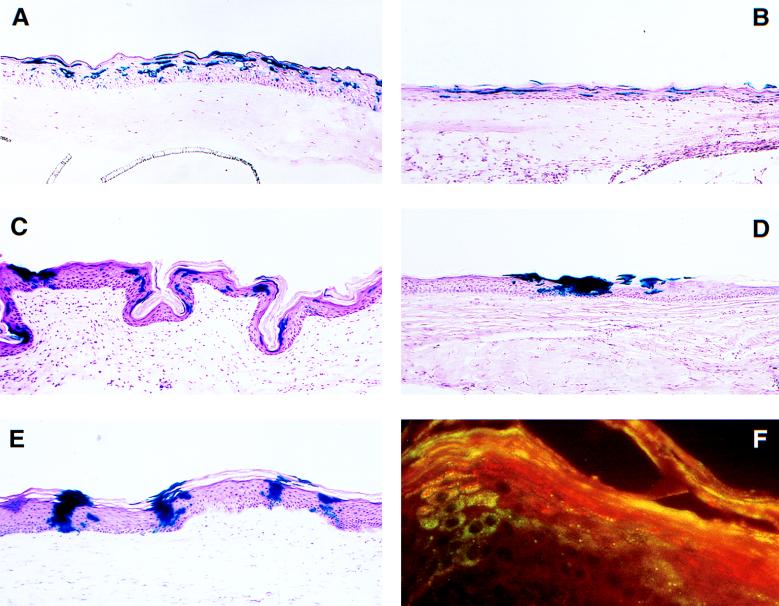
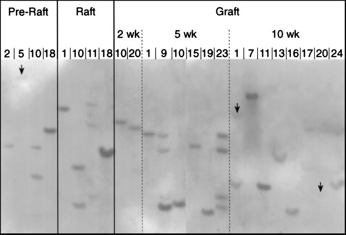
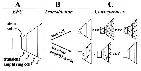
Epidermis is renewed by a population of stem cells that have been defined in vivo by slow turnover, label retention, position in the epidermis, and enrichment in beta1 integrin, and in vitro by clonogenic growth, prolonged serial passage, and rapid adherence to extracellular matrix. The goal of this study is to determine whether clonogenic cells with long-term growth potential in vitro persist in vivo and give rise to a fully differentiated epidermis. Human keratinocytes were genetically labeled in culture by transduction with a retrovirus encoding the lacZ gene and grafted to athymic mice. Analysis of the cultures before grafting showed that 21.1-27.8% of clonogenic cells with the capacity for >30 generations were successfully transduced. In vivo, beta-galactosidase (beta-gal) positive cells participated in the formation of a fully differentiated epithelium and were detected throughout the 40-week postgraft period, initially as loosely scattered clusters and later as distinct vertical columns. Viable cells recovered from excised grafts were seeded at clonal densities and 23.3-33.3% of the colonies thus formed were beta-gal positive. In addition, no evidence of transgene inactivation was obtained: all keratinocyte colonies recovered from grafted tissue that were beta-gal negative also lacked the lacZ transgene. These results show that cells with long-term growth properties in vitro do indeed persist in vivo and form a fully differentiated epidermis, thereby exhibiting the properties of stem cells.
Figures
References
-
- Potten C S. Int Rev Cytol. 1981;69:271–318. - PubMed
-
- Hall P A, Watt F M. Development (Cambridge, UK) 1989;106:619–633. - PubMed
-
- Bickenbach J R, Mackenzie I C. J Invest Dermatol. 1984;82:618–622. - PubMed
-
- Rheinwald J G, Green H. Cell. 1975;6:331–344. - PubMed
-
- Albers K M, Setzer R W, Taichman L B. Differentiation. 1986;31:134–140. - PubMed
Publication types
MeSH terms
Substances
Grants and funding
LinkOut - more resources
Full Text Sources
Other Literature Sources
Medical
