. 1998 Apr 14;95(8):4463-8.
doi: 10.1073/pnas.95.8.4463.

The lethal mutation of the mouse wasted (wst) is a deletion that abolishes expression of a tissue-specific isoform of translation elongation factor 1alpha, encoded by the Eef1a2 gene

Affiliations

The lethal mutation of the mouse wasted (wst) is a deletion that abolishes expression of a tissue-specific isoform of translation elongation factor 1alpha, encoded by the Eef1a2 gene

D M Chambers et al. Proc Natl Acad Sci U S A. .

Abstract

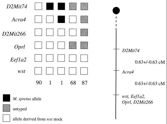
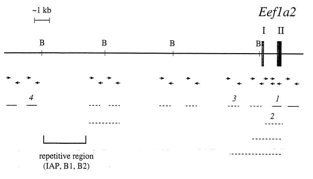
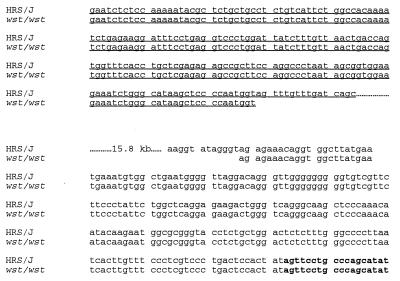
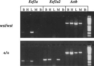
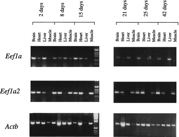
We have identified the mutation responsible for the autosomal recessive wasted (wst) mutation of the mouse. Wasted mice are characterized by wasting and neurological and immunological abnormalities starting at 21 days after birth; they die by 28 days. A deletion of 15.8 kb in wasted mice abolishes expression of a gene called Eef1a2, encoding a protein that is 92% identical at the amino acid level to the translation elongation factor EF1alpha (locus Eef1a). We have found no evidence for the involvement of another gene in this deletion. Expression of Eef1a2 is reciprocal with that of Eef1a. Expression of Eef1a2 takes over from Eef1a in heart and muscle at precisely the time at which the wasted phenotype becomes manifest. These data suggest that there are tissue-specific forms of the translation elongation apparatus essential for postnatal survival in the mouse.


Figures

Figure 1
Figure 1
Pedigree analysis of N2 offspring in the wasted backcross. Briefly, Ra+/+wst female mice were crossed to M. spretus males. Wild-type female offspring from this cross, presumptive +/wst heterozygotes, then were backcrossed to Ra+/+wst male mice. Only those offspring identified as wst/wst homozygotes were selected for further analysis. Two of the recombinants between wst and D2Mit266 listed in Abbott et al. (3) now have been shown to have been misclassified as wst/wst and have been eliminated from further analysis. The loci are listed to the left of the boxes, and the numbers below the boxes indicate the number of N2 offspring carrying that haplotype. The chromosomes shown were inherited from the F1 parent. To the right of the figure is the map derived from the haplotype analysis, giving the distances between loci as centiMorgans +/− standard error. The figure given for the distance between D2Mit74 and Acra4 is based on a total number of 166 animals scored because all of these mice were typed for the proximal marker D2Mit73 also (3).
Figure 2
Figure 2
Analysis of the region upstream of the Eef1a2 gene. BamHI restriction sites are marked “B.” Exons I and II of Eef1a2 are indicated as filled boxes. The lines represent the PCR product obtained with the primers listed below (according to the number above the line). Solid lines represent products that were obtained from both HRS/J and wst/wst DNA and dotted lines represent those that were produced only from HRS/J DNA and gave no product from wst/wst DNA. The extent of the deletion is indicated by arrows. PCR primers were as follows: product 1, 1f: 5′-TGTGGCTGAATGGGGTTAGG, 1r: 5′-CACTGTGGGGGCTCTGGTTT (product size 226 bp); product 2, 2f: 5′-CAGAGCTTCACTCAGTCTG,1r: as above (product size 379 bp); product 3, 3f: 5′-TAGTGGCTCCTTGGAACAG, 3r: 5′-CTACTCTCCCTGAATGCCTT (product size 456 bp); and product 4, 4f: 5′-TGACTATCCCATTGCCAGG, 4r: 5′-CTGCAACTCAACAGCCCATT (product size 191 bp).
Figure 3
Figure 3
Sequence analysis across the deletion breakpoint region in wasted and wild-type (HRS/J) mice. Underlined sequence represents the gag gene of the IAP element and nonunderlined sequence is intron 1 of Eef1a2. Sequence in bold is exon II of Eef1a2. Dotted lines indicate the 15.8 kb deleted in wst/wst mice.
Figure 4
Figure 4
RT-PCR of RNA from age-matched 21-day-old homozygous wst/wst and wild-type (+/+) mice. Expression of Eef1a, Eef1a2, and a β-actin control are shown. The tissues assayed are brain (B), heart (H), liver (L), and skeletal muscle (M). The RT-negative control is shown for brain only (B-); similar controls were done for all tissues, and all were completely negative. The molecular weight marker is the 1-kb ladder (GIBCO/BRL).
Figure 5
Figure 5
RT-PCR of RNA from +/+ mice of different ages, from 2 to 42 days after birth. Expression of Eef1a, Eef1a2, and a β-actin control are shown. No-RT controls were negative in all cases. The molecular weight marker is the 1-kb ladder (GIBCO/BRL).

References

    1. Shultz L D, Sweet H O, Davisson M T, Coman D R. Nature (London) 1984;297:402–404. - PubMed
    1. Lutsep H, Rodriguez M. J Neuropathol Exp Neurol. 1989;48:519–533. - PubMed
    1. Abbott C, Malas S, Pilz A, Pate L, Ali R, Peters J. Genomics. 1994;20:94–98. - PubMed
    1. Inoue T, Aikawa K, Tezuka H, Kada T, Shultz L D. Cancer Res. 1986;46:3979–3982. - PubMed
    1. Nordeen S, Schaefer V, Edgell M, Hutchison C, III, Shultz L, Swift M. Mutat Res. 1984;140:219–222. - PubMed

MeSH terms

Substances

Associated data

LinkOut - more resources