

T cell positive selection by a high density, low affinity ligand
- PMID: 9539770
- PMCID: PMC22522
- DOI: 10.1073/pnas.95.8.4522
T cell positive selection by a high density, low affinity ligand
Abstract
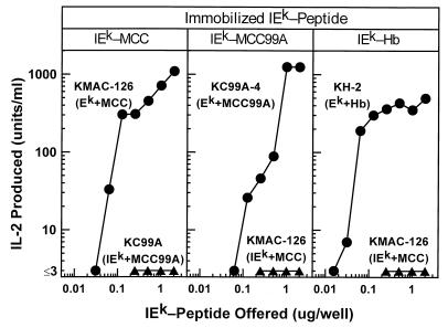
Interaction of the alpha beta T cell receptor (TCR) with major histocompatibility (MHC) molecules occupied with any of a large collection of peptides derived from self proteins is a critical step in driving T cell "positive" selection in the thymus. Interaction with this same pool of self-peptide/MHC ligands deletes T cells with potential self-reactivity. To examine how T cells survive both of these processes to form a self-tolerant mature repertoire, mice were constructed whose entire class II MHC IEk specific repertoire was positively selected on a single peptide covalently attached to the IEk molecule. In these mice T cells were identified that could respond to a variant of the positively selecting peptide bound to IEk. The affinities of the TCRs from these T cells for the positively selecting ligand were extremely low and at least 10-fold less than those for the activating ligand. These results support the theory that positive selection is driven by TCR affinities lower than those involved in T cell deletion or activation and that, if present at high concentration, even very low affinity ligands can positively select.
Figures
Similar articles
-
Selection of antigen-specific T cells by a single IEk peptide combination.J Exp Med. 1997 Nov 3;186(9):1441-50. doi: 10.1084/jem.186.9.1441. J Exp Med. 1997. PMID: 9348301 Free PMC article.
-
Alpha beta TCRs differ in the degree of their specificity for the positively selecting MHC/peptide ligand.J Immunol. 2001 Feb 15;166(4):2251-9. doi: 10.4049/jimmunol.166.4.2251. J Immunol. 2001. PMID: 11160279
-
Highly restricted T cell repertoire shaped by a single major histocompatibility complex-peptide ligand in the presence of a single rearranged T cell receptor beta chain.J Exp Med. 1998 Sep 7;188(5):897-907. doi: 10.1084/jem.188.5.897. J Exp Med. 1998. PMID: 9730891 Free PMC article.
-
Structure-function studies of T-cell receptor-superantigen interactions.Immunol Rev. 1998 Jun;163:177-86. doi: 10.1111/j.1600-065x.1998.tb01196.x. Immunol Rev. 1998. PMID: 9700510 Review.
-
Structural basis of T cell recognition of peptides bound to MHC molecules.Mol Immunol. 2002 May;38(14):1039-49. doi: 10.1016/s0161-5890(02)00033-0. Mol Immunol. 2002. PMID: 11955596 Review.
Cited by
-
Foxp1 is an essential transcriptional regulator for the generation of quiescent naive T cells during thymocyte development.Blood. 2010 Jan 21;115(3):510-8. doi: 10.1182/blood-2009-07-232694. Epub 2009 Nov 12. Blood. 2010. PMID: 19965654 Free PMC article.
-
Affinity threshold for thymic selection through a T-cell receptor-co-receptor zipper.Nat Rev Immunol. 2009 Mar;9(3):207-13. doi: 10.1038/nri2469. Nat Rev Immunol. 2009. PMID: 19151748 Review.
-
B Cell Receptor Affinity for Insulin Dictates Autoantigen Acquisition and B Cell Functionality in Autoimmune Diabetes.J Clin Med. 2016 Nov 8;5(11):98. doi: 10.3390/jcm5110098. J Clin Med. 2016. PMID: 27834793 Free PMC article.
-
A kinetic threshold between negative and positive selection based on the longevity of the T cell receptor-ligand complex.J Exp Med. 1999 May 17;189(10):1531-44. doi: 10.1084/jem.189.10.1531. J Exp Med. 1999. PMID: 10330432 Free PMC article.
-
Defining the parameters necessary for T-cell recognition of ligands that vary in potency.Immunol Res. 2004;29(1-3):29-40. doi: 10.1385/IR:29:1-3:029. Immunol Res. 2004. PMID: 15181268 Review.
References
-
- Bevan M J. Nature (London) 1977;269:417–418. - PubMed
-
- Hogquist K A, Jameson C S, Heath W R, Howard J L, Bevan M J, Carbone F R. Cell. 1994;76:17–27. - PubMed
-
- Ashton-Rickardt P G, Bandiera A, Delaney J R, van Kaer L, Pircher H P, Zinkernagel R M, Tonegawa S. Cell. 1994;76:651–663. - PubMed
-
- Sebzda E, Wallace V A, Mayer J, Yeung R S M, Mak T W, Ohashi P S. Science. 1994;263:1615–1618. - PubMed
Publication types
MeSH terms
Substances
Grants and funding
LinkOut - more resources
Full Text Sources
Research Materials
