. 1998 Apr 14;95(8):4567-72.
doi: 10.1073/pnas.95.8.4567.

Activating mutations in the extracellular domain of the fibroblast growth factor receptor 2 function by disruption of the disulfide bond in the third immunoglobulin-like domain

Affiliations

Activating mutations in the extracellular domain of the fibroblast growth factor receptor 2 function by disruption of the disulfide bond in the third immunoglobulin-like domain

S C Robertson et al. Proc Natl Acad Sci U S A. .

Abstract

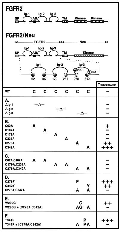
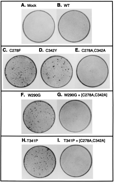
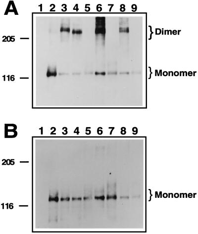
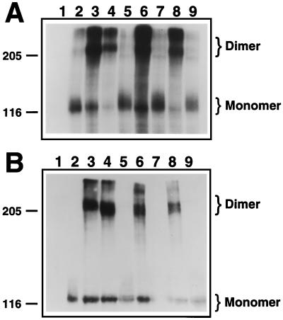
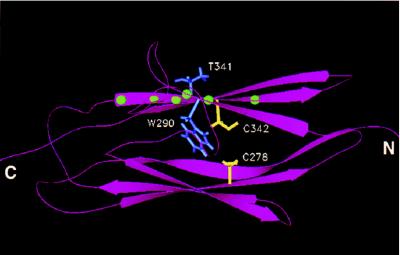
Multiple human skeletal and craniosynostosis disorders, including Crouzon, Pfeiffer, Jackson-Weiss, and Apert syndromes, result from numerous point mutations in the extracellular region of fibroblast growth factor receptor 2 (FGFR2). Many of these mutations create a free cysteine residue that potentially leads to abnormal disulfide bond formation and receptor activation; however, for noncysteine mutations, the mechanism of receptor activation remains unclear. We examined the effect of two of these mutations, W290G and T341P, on receptor dimerization and activation. These mutations resulted in cellular transformation when expressed as FGFR2/Neu chimeric receptors. Additionally, in full-length FGFR2, the mutations induced receptor dimerization and elevated levels of tyrosine kinase activity. Interestingly, transformation by the chimeric receptors, dimerization, and enhanced kinase activity were all abolished if either the W290G or the T341P mutation was expressed in conjunction with mutations that eliminate the disulfide bond in the third immunoglobulin-like domain (Ig-3). These results demonstrate a requirement for the Ig-3 cysteine residues in the activation of FGFR2 by noncysteine mutations. Molecular modeling also reveals that noncysteine mutations may activate FGFR2 by altering the conformation of the Ig-3 domain near the disulfide bond, preventing the formation of an intramolecular bond. This allows the unbonded cysteine residues to participate in intermolecular disulfide bonding, resulting in constitutive activation of the receptor.


Figures

Figure 1
Figure 1
Structure and transforming activity of FGFR2 constructs. FGFR2 is shown in comparison with FGFR2/Neu chimeric receptors, which contain the extracellular domain of FGFR2 fused to the transmembrane and intracellular domains of Neu. The six cysteine residues in the FGFR2 extracellular domain involved in intramolecular disulfide bonding are Cys-62, Cys-107, Cys-179, Cys-231, Cys-278, and Cys-342. (A) Deletions of the Ig domains remove the sequence between the disulfide bonded cysteines and replace those cysteine residues with alanine. (B) Single mutations of Cys → Ala in the extracellular domain. (C) Pair-wise mutations of Cys → Ala affecting each disulfide bond in Ig-1, Ig-2 and Ig-3. (D) Craniosynostosis mutations affecting Cys-278 and Cys-342. (E) The noncysteine craniosynostosis mutation W290G. (F) The noncysteine craniosynostosis mutation T341P. Transforming activity for each chimeric receptor, shown at right, was measured by focus formation assays in NIH 3T3 cells. Results presented are the average of at least three experiments. The results are given as a percentage of the transformation efficiency of oncogenic Neu and were reported as follows: −, 0–5%; +, 5–10%; ++, 10–20%; +++, >20%.
Figure 2
Figure 2
Transformation of NIH 3T3 cells by FGFR2/Neu chimeras. Representative plates from transformation assays described in Fig. 1 were fixed and stained with Giemsa. (A) Mock-transfected cells. (B) Wild type. (C) C278F. (D) C342Y. (E) (C278A, C342A). (F) W290G. (G) (W290G, C278A, C342A). (H) T341P. (I) (T341P, C278A, C342A).
Figure 3
Figure 3
Dimerization of activated FGFR2 mutants. COS-1 cells transiently transfected with wild-type or mutant FGFR2 expression constructs were lysed and cellular proteins were resolved on a SDS/PAGE gel. Proteins were transferred to nitrocellulose and subjected to Western blotting using antisera indicated below. (A) Samples were resolved on a 4–12% gradient gel under nonreducing conditions, incubated with anti-FGFR2 antiserum, and visualized by using enhanced chemiluminescence. Monomeric FGFR2 with an apparent molecular mass of 110 kDa and dimeric FGFR2 of approximately 220 kDa are indicated. Molecular mass markers in kDa are indicated. (B) Equivalent samples from A were resolved under reducing conditions on a 4–12% SDS/PAGE gel and visualized as described above. Lanes: 1, mock-transfected cells; 2, wild type; 3, C278F; 4, C342Y; 5, (C278A, C342A); 6, W290G; 7, (W290G, C278A, C342A); 8, T341P; 9, (T341P, C278A, C342A).
Figure 4
Figure 4
In vitro kinase assay of FGFR2 receptors. Constructs encoding FGFR2 wild-type or mutant receptors were transiently transfected into COS-1 cells. The cells were lysed, material was immunoprecipitated with FGFR2 antiserum, and an in vitro autophosphorylation reaction was performed in the presence of radiolabeled ATP. (A) Radiolabeled proteins were resolved on a SDS/PAGE 4–12% gradient gel and detected by autoradiography. (B) Identical aliquots were electrophoresed on a SDS/PAGE 4–12% gradient gel, after which proteins were transferred to nitrocellulose, subjected to immunoblotting using the anti-phosphotyrosine antiserum 4G10, and visualized via enhanced chemiluminescence. Monomeric and dimeric species of FGFR2 are indicated. Molecular mass markers in kDa are also indicated. Lanes: 1, mock-transfected cells; 2, wild type; 3, C278F; 4, C342Y; 5, (C278A, C342A); 6, W290G; 7, (W290G, C278A, C342A); 8, T341P; 9, (T341P, C278A, C342A).
Figure 5
Figure 5
Molecular modeling of Ig-3 domain of FGFR2. Molecular modeling was used to create a representation of Ig-3 of wild-type FGFR2 based on the crystallographic coordinates of the myosin light chain kinase homolog telokin. A ribbon diagram of the modeled structure is shown indicating the position of the Ig-3 cysteine residues (shown in yellow) relative to the amino acid side chains of W290 and T341 (shown in blue). The mutations W290G and T341P were examined in this study. Balls (shown in green) on the ribbon diagram indicate the positions of other noncysteine craniosynostosis mutations in the Ig-3 domain (Table 1C).

References

    1. Johnson D E, Williams L T. Adv Cancer Res. 1993;60:1–41. - PubMed
    1. Partanen J, Vainikka S, Alitalo K. Philos Trans R Soc London Ser B. 1993;340:297–303. - PubMed
    1. Ullrich A, Schlessinger J. Cell. 1990;61:203–212. - PubMed
    1. Spivak-Kroizman T, Lemmon M A, Dikic I, Ladbury J E, Pinchasi D, Huang J, Jaye M, Crumley G, Schlessinger J, Lax I. Cell. 1994;79:1015–1024. - PubMed
    1. Mohammadi M, Dikic I, Sorokin A, Burgess W H, Jaye M, Schlessinger J. Mol Cell Biol. 1996;16:977–989. - PMC - PubMed

Publication types

MeSH terms

Associated data