. 1998 Apr 14;95(8):4573-8.
doi: 10.1073/pnas.95.8.4573.

Identification and characterization of the ARP1 gene, a target for the human acute leukemia ALL1 gene

Affiliations

Identification and characterization of the ARP1 gene, a target for the human acute leukemia ALL1 gene

H Arakawa et al. Proc Natl Acad Sci U S A. .

Abstract

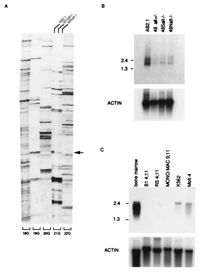
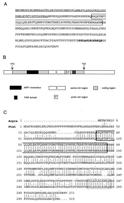
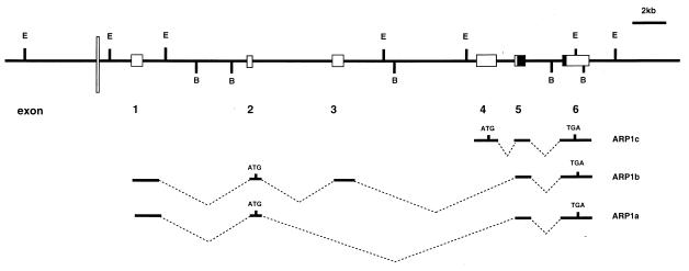
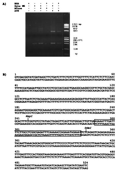
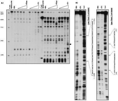
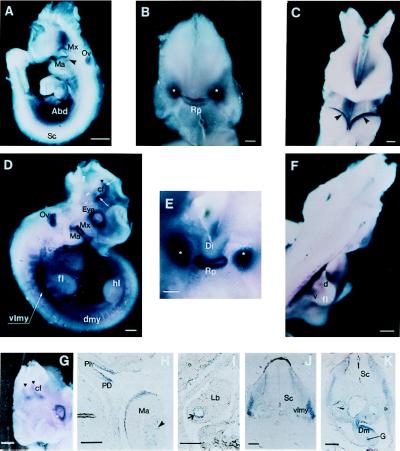
ALL1, the human homologue of Drosophila trithorax, is directly involved in human acute leukemias associated with abnormalities at 11q23. Using the differential display method, we isolated a gene that is down-regulated in All1 double-knockout mouse embryonic stem (ES) cells. The gene, designated ARP1 (also termed RIEG, Ptx2, or Otlx2), is a member of a family of homeotic genes containing a short motif shared with several homeobox genes. Using a bacterially synthesized All1 polypeptide encompassing the AT-hook motifs, we identified a 0.5-kb ARP1 DNA fragment that preferentially bound to the polypeptide. Within this DNA, a region of approximately 100 bp was protected by the polypeptide from digestion with ExoIII and DNase I. Whole-mount in situ hybridization to early mouse embryos of 9.5-10.5 days indicated a complex pattern of Arp1 expression spatially overlapping with the expression of All1. Although the ARP1 gene is expressed strongly in bone marrow cells, no transcripts were detected in six leukemia cell lines with 11q23 translocations. These results suggest that ARP1 is up-regulated by the All1 protein, possibly through direct interaction with an upstream DNA sequence of the former. The results are also consistent with the suggestion that ALL1 chimeric proteins resulting from 11q23 abnormalities act in a dominant negative fashion.


Figures

Figure 1
Figure 1
Identification of a transcript down-regulated in All1 knockout ES cells. (A) Differential display. 18G, 19G, 20G, 21G, and 22G indicate specific primer sets. The three lanes for each set correspond to the ES cell line from which the RNA was amplified: AB2.1 wild type (left lane), 48Sall−/− (center lane), and 48Nall−/− (right lane). Arrow points to the band apparent only in the wild-type cells. (B) Northern analysis of Arp1 expression in ES cell lines. Two micrograms of polyadenylated RNA were electrophoretically resolved and examined for hybridization to the 161-bp probe (see text). (C) Northern analysis of ARP1 expression in human leukemia cell lines. Analysis as in B. The 300-bp human cDNA (see text) was utilized as a probe. Numbers on the left correspond to size markers (kb).
Figure 2
Figure 2
Amino acids sequence, domains, and homology of ARP1. (A) Amino acids sequence of ARP1c. Underline indicates unique domain of ARP1c; box indicates homeodomain. Bold letters are short motifs mentioned by Semina et al. (28). (B) Schema of domain of ARP1c. (C) Similarity between the Arp1a and Ptx1 proteins. The sequences were aligned by using the algorithm bestfit.
Figure 3
Figure 3
Genomic structure of human ARP1. Exons are depicted by lightly shaded boxes. Solid box indicates ARP1 homeodomain. Box sizes do not correspond to precise length of exons. The All1 AT-hook binding site is noted by thin, white bar. E and B correspond to EcoRI and BamHI sites, respectively. Exons included within ARP1 transcripts are shown in the middle.
Figure 4
Figure 4
Binding-site selection of ARP1 genomic fragments. (A) Bacterially synthesized All1 polypeptides spanning the “AT-hooks” domain (ATH) or the region homologous to methyltransferases (MTase), together or without BSA or HeLa nuclear extract (NE), were reacted with the fragmented ARP1 genomic DNA. Bound fragments were PCR-amplified and identified by agarose gel electrophoresis. (B) The sequence of the ATH-selected DNA fragment. Sequence protected by ATH from ExoIII digestive is boxed.
Figure 5
Figure 5
Fine mapping of ARP1 genomic sequence that binds the ATH polypeptide. (A Left and Right) Protection from ExoIII digestion of ARP1 DNA segment end-labeled at the 5′ and 3′ ends, respectively. Bands protected specifically by increasing amounts of the ATH polypeptide are indicated by arrows. (B) Protection from DNaseI digestion of ARP1 DNA segment labeled at the 5′ or 3′ end.
Figure 6
Figure 6
Spatial distribution of Arp1 transcripts during mouse development. (A) Lateral view of E9.5 embryo. Hybridizable RNA is observed in the abdominal area (Abd), in surface ectoderm of the maxillary (Mx), and mandibular (Ma) regions of the first branchial arch. Arrowhead points out the mesenchymal component of the mandibular arch. Staining of the optic vesicle (Ov) is because of the nonspecific trapping of colored conjugates as evidenced by reaction with a sense probe. No signal was observed in spinal cord (Sc). (B) Anterior view of the head of E9.5 embryo. Facial part has been removed for better visualization of the signal. Staining is observed in the developing Rathke’s pouch (Rp) and in two bilateral structures (marked by white asterisks), still to be identified. (C) Anterior view of the first branchial arches of E9.5 embryo. Arrowheads point to the sharp bands of Arp1 expression along the surface ectoderm of the mandibular component of the first branchial arch. (D) Lateral view of E10.5 embryo. Expression is detected in a number of new structures: in the dermomyotomal compartments of the somites (dmy), in the presumable migratory myogenic precursors (of the limb muscle) localized in the ventrolateral myotome (vlmy) at the forelimb level. The signal is also seen at the periphery of the eye. Strong staining is observed in the restricted domains of the forebrain (white arrows) at the area of the cranial flexure (cf). Black triangle corresponds to the midbrain/forebrain boundary; fl, forelimb; hl, hindlimb. (E) High magnification of the anterior view of head of E10.5 embryo (facial part has been removed); expression of Arp1 is sharply localized to the Rathke’s pouch. Bilateral staining previously detected becomes stronger and more compact. (F) Dorsolateral view of E10.5 embryo. Arp1 specific signal is seen in presumable ventral (v) and dorsal (d) myoblast cells within the forelimb (fl). (G) Anteriolateral view of E10.5 head showing bilateral distribution of Arp1 signal in the forebrain. (H) Sagittal section of the prestained E10.5 embryo. Expression is stronger in the pars dislis (PD) than in pars intermedia (PI) within the Rathke’s pouch. Expression is seen in the ectoderm and mesenchyme (arrowhead) of the mandibular branch (Ma) of the first branchial arch. (I) Staining was detected on sagittal section in epithelial cells of the bronchi (arrow) within the lung bud (Lb). (J and K) Transverse sections of the E10.5 embryo at the level of the forelimbs. Open triangle points to the myoblast precursor cells migrating into the proximal portion of the forelimb. Staining is also revealed in the mesenchyme of the dorsal mesentery (Dm); G, gut. [Bars = 200 mm (A, D, and FI), 100 mm (J and K), and 50 mm (B and C).]

References

    1. Gu Y, Nakamura T, Alder H, Prasad R, Canaani O, Cimino G, Croce C M, Canaani E. Cell. 1992;71:701–708. - PubMed
    1. Tkachuk D C, Kohler S, Cleary M L. Cell. 1992;71:691–700. - PubMed
    1. Mazo A M, Huang D H, Mozer B A, David I B. Proc Natl Acad Sci USA. 1990;87:2112–2116. - PMC - PubMed
    1. Ma Q, Alder H, Nelson K K, Chatterjel D, Gu Y, Nakamura T, Canaani E, Croce C M, Siracusa L D, Buchberg A M. Proc Natl Acad Sci USA. 1993;90:6350–6354. - PMC - PubMed
    1. Zeleznick-Le N J, Harden A M, Rowley J D. Proc Natl Acad Sci USA. 1994;91:10610–10614. - PMC - PubMed

Publication types

MeSH terms