Case Reports
. 1998 Apr 14;95(8):4584-8.
doi: 10.1073/pnas.95.8.4584.

Fetal origins of the TEL-AML1 fusion gene in identical twins with leukemia

Affiliations
Case Reports

Fetal origins of the TEL-AML1 fusion gene in identical twins with leukemia

A M Ford et al. Proc Natl Acad Sci U S A. .

Abstract

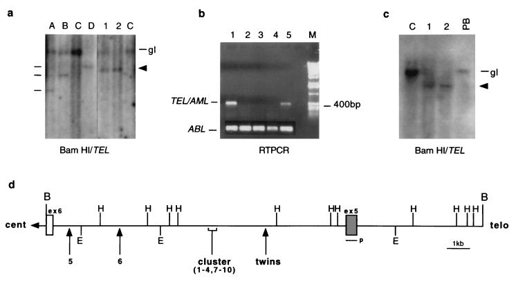
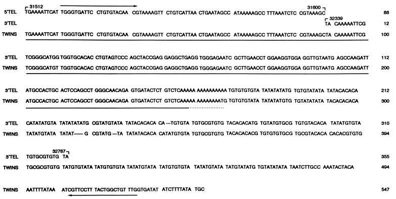
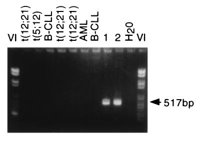
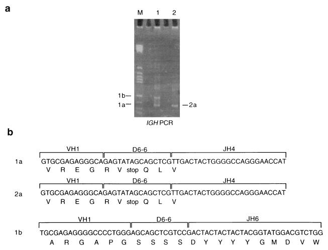
The TEL (ETV6)-AML1 (CBFA2) gene fusion is the most common reciprocal chromosomal rearrangement in childhood cancer occurring in approximately 25% of the most predominant subtype of leukemia- common acute lymphoblastic leukemia. The TEL-AML1 genomic sequence has been characterized in a pair of monozygotic twins diagnosed at ages 3 years, 6 months and 4 years, 10 months with common acute lymphoblastic leukemia. The twin leukemic DNA shared the same unique (or clonotypic) but nonconstitutive TEL-AML1 fusion sequence. The most plausible explanation for this finding is a single cell origin of the TEL-AML fusion in one fetus in utero, probably as a leukemia-initiating mutation, followed by intraplacental metastasis of clonal progeny to the other twin. Clonal identity is further supported by the finding that the leukemic cells in the two twins shared an identical rearranged IGH allele. These data have implications for the etiology and natural history of childhood leukemia.


Figures

Figure 1
Figure 1
Molecular analysis of t(12;21) in identical twins. (a) BamHI Southern blot analysis of TEL rearrangement. The monozygotic twins, lanes 1 and 2, respectively, show a shared pattern of TEL rearrangement (indicated by arrow) and loss of heterozygosity. Lanes A, B, and D are t(12;21) controls whose different rearrangements are denoted by a line. Lanes C represent AML controls, and gl is germ line (20 kb). (b) RT-PCR analysis for TEL-AML fusion transcript. Lanes 2–4, childhood ALL cases negative for t(12;21). Lane 1, twin 1 (bone marrow at diagnosis 59% blasts), lane 5 twin 2 (bone marrow at diagnosis 97% blasts). (Lower) ABL RT-PCR. (c) The TEL rearrangement is nonconstitutive. BamHI Southern blot analysis showing a lack of TEL rearrangements in diagnostic peripheral blood of twin 1 (PB; 2% blasts). C is the AML control, 1 and 2 represent each twin, and gl is germ-line 20 kb. The rearranged TEL allele is indicated by an arrow. (d) Partial restriction map of the TEL breakpoint cluster region. The map shows only the relevant restriction sites and is adapted from ref. . B, BamHI; E, EcoRI; H, HindIII; p, probe. The location of the TEL breakpoint in the twins is indicated by an arrow in relation to 10 previously published breaks (9). Cent relates to centromere and telo to telomere.
Figure 2
Figure 2
Sequence comparison between TEL intron 5 and twin 1 breakpoint sequences. 5′ TEL refers to published intron 5 sequence from 31512 to 31600 (14). 3′ TEL refers to sequences 32339 to 32787. The twins sequence is shown underneath with a 739-bp deletion between the 5′ and 3′ TEL sequences. Twin sequence diverges from TEL at 32787 presumably into AML1 intron sequence. The sequence with ≈100% homology to a human carcinoma cell-derived Alu RNA transcript (NE36, ref. 24) is underlined. Arrows indicate PCR primers.
Figure 3
Figure 3
The TEL/AML1 genomic breakpoint is identical in each twin. Breakpoint-specific PCR primers were used to amplify the fusions from a number of patient DNAs with and without t(12;21). Only DNA from the twins gives the expected 517-bp product showing breakpoint sequence specificity.
Figure 4
Figure 4
An IGH VDJ join is identical in each twin. (a) PAGE of PCR product from IGH gene rearrangements in the twin pairs showing two identically sized fragments and one additional band specific to twin 1. All bands were cut out, cloned, and sequenced. (b) Nucleotide and amino acid composition of the IGH products. VH, variable region; DH, diversity region; JH, joining region. V, D, and J motifs were identified from refs. –.

References

    1. Sawyers C L. Lancet. 1997;349:196–200. - PubMed
    1. Hagemeijer A, Grosveld G. In: Leukemia. Henderson E S, Lister T A, Greaves M F, editors. Philadelphia: Saunders; 1996. pp. 131–144.
    1. Rabbitts T H. Nature (London) 1994;372:143–149. - PubMed
    1. Look A T. Science. 1997;278:1059–1064. - PubMed
    1. Golub T R, Barker G F, Stegmaier K, Gilliland D G. Curr Top Microbiol Immunol. 1996;211:279–288. - PubMed

Publication types

MeSH terms

Substances

Associated data