. 1998 Apr 14;95(8):4607-12.
doi: 10.1073/pnas.95.8.4607.

Regulation of transport pathways in tumor vessels: role of tumor type and microenvironment

Affiliations

Regulation of transport pathways in tumor vessels: role of tumor type and microenvironment

S K Hobbs et al. Proc Natl Acad Sci U S A. .

Abstract

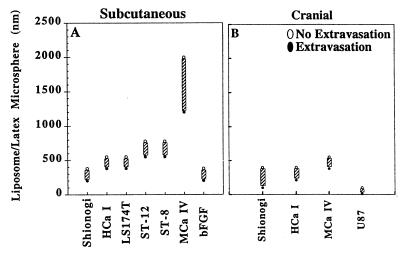
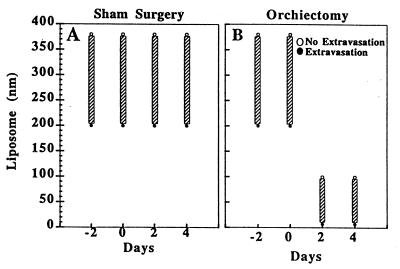
Novel anti-neoplastic agents such as gene targeting vectors and encapsulated carriers are quite large (approximately 100-300 nm in diameter). An understanding of the functional size and physiological regulation of transvascular pathways is necessary to optimize delivery of these agents. Here we analyze the functional limits of transvascular transport and its modulation by the microenvironment. One human and five murine tumors including mammary and colorectal carcinomas, hepatoma, glioma, and sarcoma were implanted in the dorsal skin-fold chamber or cranial window, and the pore cutoff size, a functional measure of transvascular gap size, was determined. The microenvironment was modulated: (i) spatially, by growing tumors in subcutaneous or cranial locations and (ii) temporally, by inducing vascular regression in hormone-dependent tumors. Tumors grown subcutaneously exhibited a characteristic pore cutoff size ranging from 200 nm to 1.2 microm. This pore cutoff size was reduced in tumors grown in the cranium or in regressing tumors after hormone withdrawal. Vessels induced in basic fibroblast growth factor-containing gels had a pore cutoff size of 200 nm. Albumin permeability was independent of pore cutoff size. These results have three major implications for the delivery of therapeutic agents: (i) delivery may be less efficient in cranial tumors than in subcutaneous tumors, (ii) delivery may be reduced during tumor regression induced by hormonal ablation, and (iii) permeability to a molecule is independent of pore cutoff size as long as the diameter of the molecule is much less than the pore diameter.


Figures

Figure 1
Figure 1
The vascular pore cutoff size for six different types of tumors grown in the dorsal chamber (A) and four tumors grown in the cranial window (B) was evaluated. The solid circles represent significant extravasation at the indicated long-circulating liposome or latex bead size. The open circles represent no extravasation at the indicated liposome/latex bead size. The size range between the last particle extravasated and the first particle that did not extravasate indicates the vascular pore cutoff size range (hatched bar). The majority of tumors have a vascular pore cutoff size range between 380 and 780 nm when grown subcutaneously in the dorsal chamber. The interaction of the tumor with the cranial microenvironment (B) leads to a smaller vascular pore cutoff size than the interaction of the same tumor with the subcutaneous microenvironment (A). Comparison of bFGF-induced vessels (bFGF) and tumor-induced vascular pore sizes demonstrates that the presence of bFGF alone can lead to pores of this size.
Figure 2
Figure 2
Representative electron micrographs of MCa IV tumor vessels grown in the dorsal chamber. (A) Venular-sized vessel with fenestrated endothelium (arrowheads). (Bar = 500 nm.) (B) Tumor vessel with an open gap measuring 856 nm delineated by the arrows. (Bar = 500 nm.) (C) Open endothelial gap in tumor blood vessel from a long-circulating liposome-injected animal. What appear to be long-circulating liposomes are shown traversing through the open junction. Serial sections (not shown) demonstrate that these are not transverse sections of cellular projections. (Bar = 300 nm.)
Figure 3
Figure 3
Comparison of the tumor size and vascularization during growth and regression of Shionogi xenografts in the dorsal chamber. Orchiectomy or sham operation was conducted after 11 days of tumor growth (Op Day). Tumor regression and growth is documented at postoperative day 12 for orchiectomized and sham-operated mice, respectively.
Figure 4
Figure 4
Shionogi tumors were implanted in the dorsal chamber of male severe combined immunodeficient mice, and pore cutoff size was determined during microvascular growth and regression for a maximum of 27 days. Initial vascularization occurred within 7 days. Day 0 corresponds to the date of orchiectomy or sham operation, day −2 is 48 hr before the surgery, and day 2 is 48 hr after surgery. (A) Sham-operated. Once the tumors were vascularized, there was no change in the vascular pore cutoff size with further growth of the tumor. (B) Orchiectomized. However, there was a rapid and striking decrease in the vascular pore cutoff size with hormone-withdrawal-mediated tumor regression. The vascular pore cutoff size returned to that of normal tissue within 48 hr after orchiectomy.

References

    1. Gerlowski L, Jain R. Microvasc Res. 1986;31:288–305. - PubMed
    1. Jain R. Science. 1996;271:1079–1080. - PubMed
    1. Jain R. Cancer Metastasis Rev. 1987;6:559–593. - PubMed
    1. Yuan F, Dellian M, Fukumura D, Leunig M, Berk D, Torchillin V, Jain R. Cancer Res. 1995;55:3752–3756. - PubMed
    1. Fukumura D, Yuan F, Monsky W, Chen Y, Jain R. Am J Pathol. 1997;151:679–688. - PMC - PubMed

Publication types

MeSH terms

LinkOut - more resources