. 1998 Apr 14;95(8):4613-8.
doi: 10.1073/pnas.95.8.4613.

Direct adenovirus-mediated gene transfer of interleukin 1 and tumor necrosis factor alpha soluble receptors to rabbit knees with experimental arthritis has local and distal anti-arthritic effects

Affiliations

Direct adenovirus-mediated gene transfer of interleukin 1 and tumor necrosis factor alpha soluble receptors to rabbit knees with experimental arthritis has local and distal anti-arthritic effects

S C Ghivizzani et al. Proc Natl Acad Sci U S A. .

Abstract

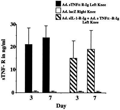
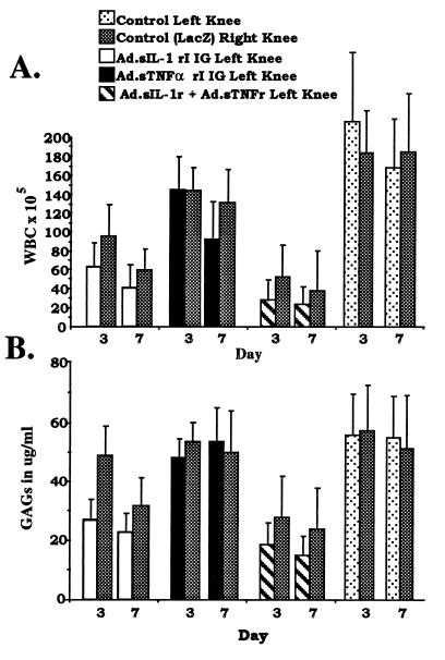
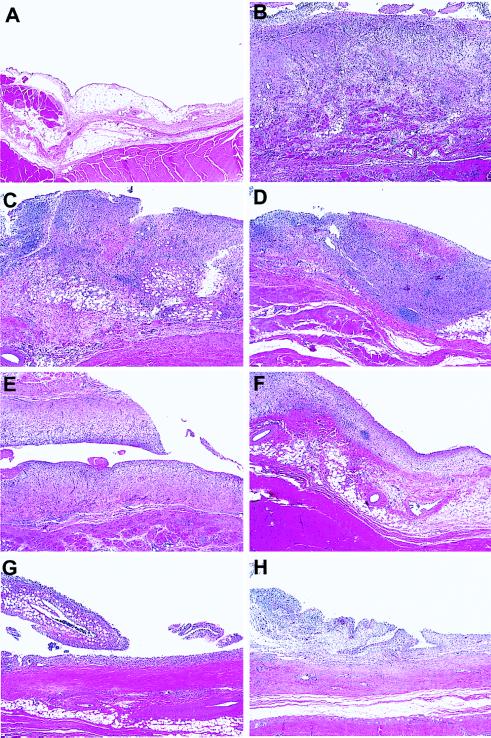
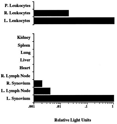
Adenoviral vectors were used to deliver genes encoding a soluble interleukin 1 (IL-1)-type I receptor-IgG fusion protein and/or a soluble type I tumor necrosis factor alpha (TNFalpha) receptor-IgG fusion protein directly to the knees of rabbits with antigen-induced arthritis. When tested individually, knees receiving the soluble IL-1 receptor had significantly reduced cartilage matrix degradation and white blood cell infiltration into the joint space. Delivery of the soluble TNFalpha receptor was less effective, having only a moderate effect on white blood cell infiltration and no effect on cartilage breakdown. When both soluble receptors were used together, there was a greater inhibition of white blood cell infiltration and cartilage breakdown with a considerable reduction of synovitis. Interestingly, anti-arthritic effects were also seen in contralateral control knees receiving only a marker gene, suggesting that sustained local inhibition of disease activity in one joint may confer an anti-arthritic effect on other joints. These results suggest that local intra-articular gene transfer could be used to treat systemic polyarticular arthritides.


Figures

Figure 1
Figure 1
In vivo expression of TNFα inhibitor after injection into rabbit knees with a.i.a. Twenty-four hours after induction of a.i.a. two groups of eight rabbits were injected in the left knee with either 7 × 10⁷ pfu of Ad.sTNF-RI-Ig or 7 × 10⁷ pfu of Ad.sTNF-RI-Ig and Ad.sIL-1-RI-Ig. Both groups were injected with 7 × 10⁷ pfu of Ad.lacZ in the right knee. At days 3 and 7 after injection of virus the knees were lavaged and the recovered fluids were analyzed by ELISA for levels of human type I TNFα soluble receptor (sTNF-R).
Figure 2
Figure 2
Leukocytic infiltration and GAG release in a.i.a. knees after intraarticular injection of Ad.sIL-1-RI-Ig and Ad.sTNF-RI-Ig. a.i.a. was induced in both knees of four groups of eight rabbits. Twenty-four hours after induction groups of rabbits received injection of adenoviral vectors as indicated. (A) At 3 and 7 days after adenoviral injection, knees of rabbits were lavaged. Numbers of infiltrating leukocytes (white blood cells; WBC) in synovial fluids for both right and left knees of each group are shown as indicated. (B) GAG levels in lavage fluids were determined by 1,9-dimethylmethylene blue assay and are shown for each group as indicated. Error bars designate ± one standard deviation. To determine whether differences observed among the groups of rabbits, left and right knees, and the days were significant, the appropriate F tests were performed. For A, differences among the groups, knees, and days, P values equal 0.0001, 0.0365, and 0.0008, respectively. For B, differences among the groups, knees, and days, P values equal 0.0001, 0.0149, and 0.0312, respectively.
Figure 3
Figure 3
Histological analyses of synovial tissue recovered from rabbit knees. a.i.a. was induced in both knees of four groups of eight rabbits. Twenty-four hours after induction all rabbits were injected in the right knee with 7 × 10⁷ pfu of Ad.lacZ. In the left knee, groups of 8 rabbits were injected with 7 × 10⁷ pfu of Ad.sTNF-RI-Ig, 7 × 10⁷ pfu of Ad.sIL-1-RI-Ig, 7 × 10⁷ pfu each of Ad.sTNF-RI-Ig and Ad.sIL-1-RI-Ig, or saline. Seven days after virus injection synovial tissues from each group were harvested, sectioned, and stained with hematoxylin and eosin. (×40.) (A) Synovium from a normal naive rabbit knee. (B) Synovium recovered from rabbit knee receiving saline after a.i.a. induction. Relative to A, the tissue is dramatically thickened, fibrous, and hypercellular, with infiltrating leukocytes and synovial cells. (C and D) Synovial tissue recovered from rabbit knees injected with Ad.sTNF-RI-Ig in the left (C) and Ad.lacZ in the right (D). (E and F) Tissue recovered from rabbit knees injected with Ad.sIL-1-RI-Ig (E) in the left and Ad.lacZ in the right (F). (G and H) Synovium from rabbits injected with both Ad.sTNF-RI-Ig and Ad.sIL-1-RI-Ig in the left knee and Ad.lacZ in the right.
Figure 4
Figure 4
Expression of luciferase activity in various tissues after intraarticular injection of Ad.luciferase into a.i.a. knees. Twenty-four hours after induction of a.i.a. in both knees of two rabbits, 1.5 × 10⁹ pfu of Ad.luciferase was injected into the left knee joint (L.), and 7 × 10⁷ pfu of Ad.lacZ was injected into the right knee joint (R.). Seven days after injection of adenovirus, the rabbits were sacrificed. Blood was collected, knees were lavaged, and synovium, heart, liver, lung, spleen, and kidney tissues were harvested. Luciferase activities in lysates from tissues and leukocytes from lavages of right (R.) and left (L.) knees and blood (peripheral [P.] leukocytes) were quantitated and are expressed relative to luciferase activity found in synovium and leukocytes from the left knee.

References

    1. Kang R, Ghivizzani S C, Herndon J H, Robbins P D, Evans C H. Biochem Soc Transact. 1997;25:533–537. - PubMed
    1. Arend W P, Dayer J M. Arthritis Rheum. 1995;38:151–160. - PubMed
    1. Feldmann M, Elliot M J, Woody J N, Maini R N. Adv Immunol. 1997;64:283–350. - PubMed
    1. Otani K, Nita I, Macaulay W, Georgescu H I, Robbins P D, Evans C H. J Immunol. 1996;156:3558–3562. - PubMed
    1. Makarov S S, Olsen J C, Johnston W N, Anderle S K, Brown R R, Baldwin A S, Jr, Haskill J S, Schwab J H. Proc Natl Acad Sci USA. 1996;93:402–406. - PMC - PubMed

Publication types

MeSH terms