

An antagonistic vascular endothelial growth factor (VEGF) variant inhibits VEGF-stimulated receptor autophosphorylation and proliferation of human endothelial cells
- PMID: 9539788
- PMCID: PMC22540
- DOI: 10.1073/pnas.95.8.4625
An antagonistic vascular endothelial growth factor (VEGF) variant inhibits VEGF-stimulated receptor autophosphorylation and proliferation of human endothelial cells
Abstract
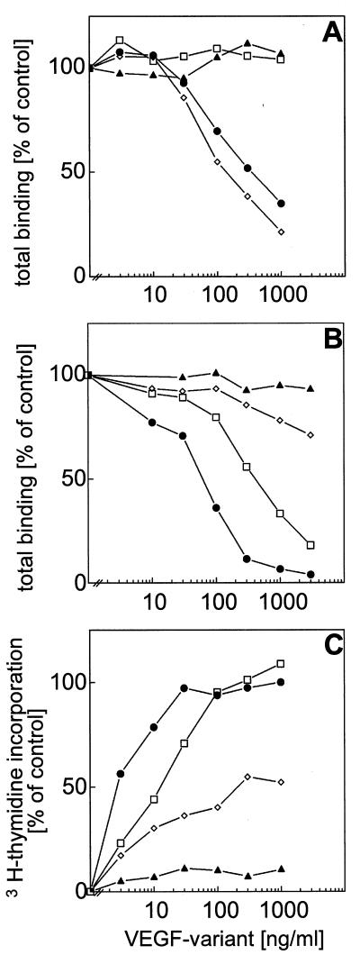
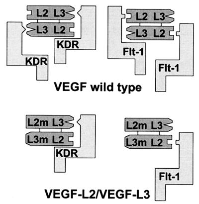
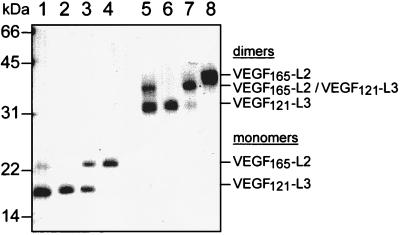
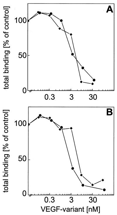
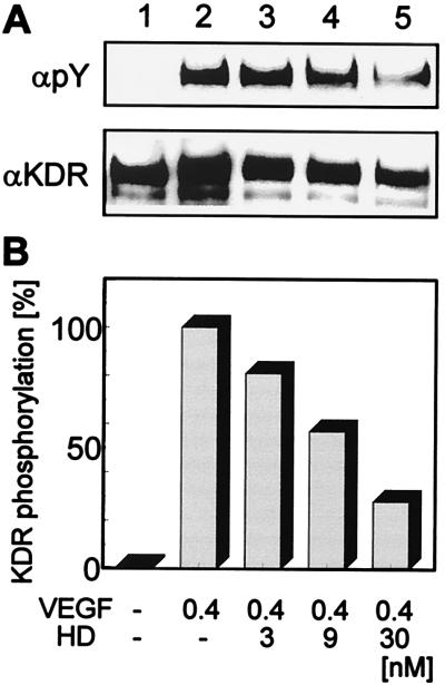
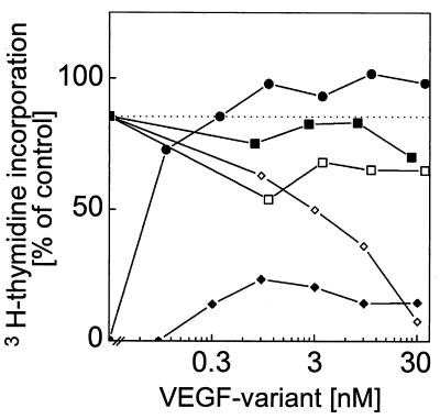
Vascular endothelial growth factor (VEGF) is a potent mitogen with a unique specificity for endothelial cells and a key mediator of aberrant endothelial cell proliferation and vascular permeability in a variety of human pathological situations, such as tumor angiogenesis, diabetic retinopathy, rheumatoid arthritis, or psoriasis. VEGF is a symmetric homodimeric molecule with two receptor binding interfaces lying on each pole of the molecule. Herein we report on the construction and recombinant expression of an asymmetric heterodimeric VEGF variant with an intact receptor binding interface at one pole and a mutant receptor binding interface at the second pole of the dimer. This VEGF variant binds to VEGF receptors but fails to induce receptor activation. In competition experiments, the heterodimeric VEGF variant antagonizes VEGF-stimulated receptor autophosphorylation and proliferation of endothelial cells. A 15-fold excess of the heterodimer was sufficient to inhibit VEGF-stimulated endothelial cell proliferation by 50%, and a 100-fold excess resulted in an almost complete inhibition. By using a rational approach that is based on the structure of VEGF, we have shown the feasibility to construct a VEGF variant that acts as an VEGF antagonist.
Figures
Similar articles
-
Expression of biologically active isoforms of the tumor angiogenesis factor VEGF in Escherichia coli.Biochem Biophys Res Commun. 1996 May 15;222(2):249-55. doi: 10.1006/bbrc.1996.0730. Biochem Biophys Res Commun. 1996. PMID: 8670191
-
Identification of vascular endothelial growth factor determinants for binding KDR and FLT-1 receptors. Generation of receptor-selective VEGF variants by site-directed mutagenesis.J Biol Chem. 1996 Mar 8;271(10):5638-46. doi: 10.1074/jbc.271.10.5638. J Biol Chem. 1996. PMID: 8621427
-
Heterodimers of placenta growth factor/vascular endothelial growth factor. Endothelial activity, tumor cell expression, and high affinity binding to Flk-1/KDR.J Biol Chem. 1996 Feb 9;271(6):3154-62. doi: 10.1074/jbc.271.6.3154. J Biol Chem. 1996. PMID: 8621715
-
Possible involvement of VEGF-FLT tyrosine kinase receptor system in normal and tumor angiogenesis.Princess Takamatsu Symp. 1994;24:162-70. Princess Takamatsu Symp. 1994. PMID: 8983073 Review.
-
Targeting tumor vasculature using VEGF-toxin conjugates.Methods Mol Biol. 2001;166:219-34. doi: 10.1385/1-59259-114-0:219. Methods Mol Biol. 2001. PMID: 11217369 Review. No abstract available.
Cited by
-
Blood vessel maturation, vascular phenotype and angiogenic potential in malignant melanoma: one step forward for overcoming anti-angiogenic drug resistance?Mol Oncol. 2011 Apr;5(2):137-49. doi: 10.1016/j.molonc.2011.01.003. Epub 2011 Feb 3. Mol Oncol. 2011. PMID: 21345752 Free PMC article. Review.
-
Dimerization of VEGF receptors and implications for signal transduction: a computational study.Biophys Chem. 2007 Jul;128(2-3):125-39. doi: 10.1016/j.bpc.2007.03.010. Epub 2007 Mar 24. Biophys Chem. 2007. PMID: 17442480 Free PMC article.
-
Inhibition of tumor growth and metastasis by targeting tumor-associated angiogenesis with antagonists to the receptors of vascular endothelial growth factor.Invest New Drugs. 1999;17(3):195-212. doi: 10.1023/a:1006314501634. Invest New Drugs. 1999. PMID: 10665474 Review.
-
Impaired in vivo vasculogenic potential of endothelial progenitor cells in comparison to human umbilical vein endothelial cells in a spheroid-based implantation model.Cell Prolif. 2009 Aug;42(4):498-505. doi: 10.1111/j.1365-2184.2009.00610.x. Epub 2009 May 29. Cell Prolif. 2009. PMID: 19489982 Free PMC article.
-
Antagonistic VEGF variants engineered to simultaneously bind to and inhibit VEGFR2 and alphavbeta3 integrin.Proc Natl Acad Sci U S A. 2011 Aug 23;108(34):14067-72. doi: 10.1073/pnas.1016635108. Epub 2011 Aug 8. Proc Natl Acad Sci U S A. 2011. PMID: 21825147 Free PMC article.
References
-
- Martiny-Baron G, Marmé D. Curr Opin Biotechnol. 1995;6:675–680. - PubMed
-
- Thomas K A. J Biol Chem. 1996;271:603–606. - PubMed
-
- Breier G, Risau W. Trends Cell Biol. 1996;6:454–456. - PubMed
-
- Risau W. Nature (London) 1997;386:671–674. - PubMed
-
- Carmeliet P, Ferreira V, Breier G, Pollefeyt S, Kieckens L, Gertsenstein M, Fahrig M, Vandenhoeck A, Harpal K, Eberhardt C, et al. Nature (London) 1996;380:435–439. - PubMed
Publication types
MeSH terms
Substances
LinkOut - more resources
Full Text Sources
Other Literature Sources
