. 1998 Apr 14;95(8):4646-51.
doi: 10.1073/pnas.95.8.4646.

NF-kappa B-dependent inhibition of apoptosis is essential for host cellsurvival during Rickettsia rickettsii infection

Affiliations

NF-kappa B-dependent inhibition of apoptosis is essential for host cellsurvival during Rickettsia rickettsii infection

D R Clifton et al. Proc Natl Acad Sci U S A. .

Abstract

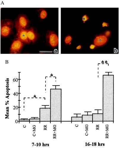
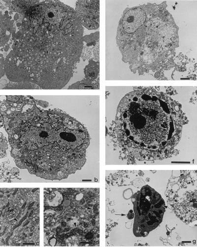
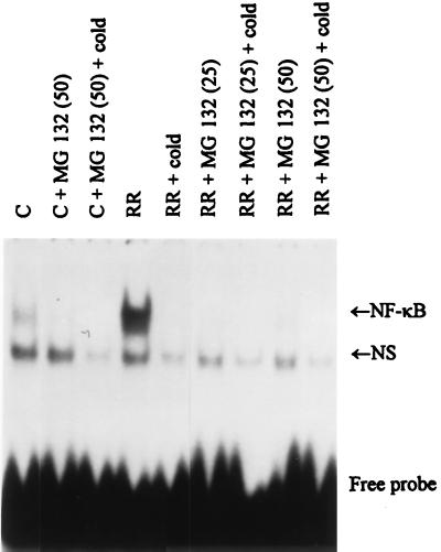
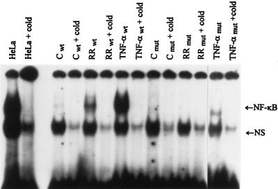
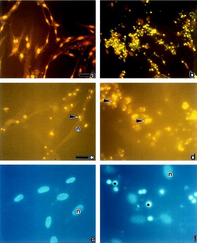
The possibility that bacteria may have evolved strategies to overcome host cell apoptosis was explored by using Rickettsia rickettsii, an obligate intracellular Gram-negative bacteria that is the etiologic agent of Rocky Mountain spotted fever. The vascular endothelial cell, the primary target cell during in vivo infection, exhibits no evidence of apoptosis during natural infection and is maintained for a sufficient time to allow replication and cell-to-cell spread prior to eventual death due to necrotic damage. Prior work in our laboratory demonstrated that R. rickettsii infection activates the transcription factor NF-kappa B and alters expression of several genes under its control. However, when R. rickettsii-induced activation of NF-kappa B was inhibited, apoptosis of infected but not uninfected endothelial cells rapidly ensued. In addition, human embryonic fibroblasts stably transfected with a superrepressor mutant inhibitory subunit Ikappa B that rendered NF-kappa B inactivatable also underwent apoptosis when infected, whereas infected wild-type human embryonic fibroblasts survived. R. rickettsii, therefore, appeared to inhibit host cell apoptosis via a mechanism dependent on NF-kappa B activation. Apoptotic nuclear changes correlated with presence of intracellular organisms and thus this previously unrecognized proapoptotic signal, masked by concomitant NF-kappa B activation, likely required intracellular infection. Our studies demonstrate that a bacterial organism can exert an antiapoptotic effect, thus modulating the host cell's apoptotic response to its own advantage by potentially allowing the host cell to remain as a site of infection.


Figures

Figure 1
Figure 1
(A) In situ detection of EC apoptosis during R. rickettsii infection. Infected human umbilical vein ECs were incubated in the presence (b) or absence (a) of the NF-κB inhibitor MG 132. At 18 hr, ECs were fixed and processed for in situ detection of DNA fragmentation by TUNEL and then counterstained with propidium iodide. When visualized under dual wavelength fluorescence, normal nuclei exhibited orange fluorescence (n), whereas apoptotic nuclei displayed green fluorescence and appeared condensed (∗). MG 132 treatment did not induce apoptosis in uninfected ECs (data not shown). (Bar = 20 μm.) (B) The percentages of apoptotic ECs were determined by scoring cells as apoptotic (exhibiting green fluorescence) or normal (exhibiting orange fluorescence) in randomly chosen microscopic fields. Fifteen hundred to 3,000 cells were scored per experimental condition. P values comparing mean percent apoptotic cells between experimental conditions indicated by brackets were determined by Student’s t test and are indicated by asterisks (n = 3 to 5). ∗, P < 0.05; ∗∗, P < 0.00005.
Figure 2
Figure 2
Transmission electron micrographs of R. rickettsii-infected ECs. Shown are uninfected ECs (a and c) or ECs infected for 38 hr in the absence (b and d) or presence (e–g) of MG 132. EC nuclei are denoted by n. Arrowheads in c and d point to mitochondria. Arrowheads in f and g point to areas of condensed chromatin; the arrow in g points to an apoptotic body. [Bars = 2.5 μm (a, b, and e–g) and 1 μm (c and d).]
Figure 3
Figure 3
Effect of MG 132 on R. rickettsii induced NF-κB activation. Gel shift analysis using a 32P-labeled oligonucleotide probe was performed on nuclear extracts prepared from control ECs (C) and ECs infected for 3 hr with R. rickettsii (RR) in the absence and presence of 25 μM (25) or 50 μM (50) MG 132. Analysis of complex formation was performed on 4% nonreducing polyacrylamide gels followed by autoradiography. An R. rickettsii-inducible gel-shifted complex representing interaction of dimeric NF-κB molecules with the labeled probe is indicated in the gel (NF-κB) as is the presence of a nonspecific noninducible gel-shifted complex (NS). Specificity of the NF-κB gel-shifted complexes was demonstrated by inclusion of excess unlabeled oligonucleotide probe (+cold).
Figure 4
Figure 4
Gel shift analysis of NF-κB induction in HEF cells. Gel shift analysis was performed on nuclear extracts prepared from control (C), R. rickettsii-infected (RR), and TNF-α-treated (10 ng/ml) wild-type (wt) and mutant (mut) HEFs incubated for 3 hr. Complexes were analyzed on 4% nondenaturing polyacrylamide gels followed by autoradiography. HeLa cells were included as positive markers. Other symbols are as in Fig. 3.
Figure 5
Figure 5
Induction of apoptosis in wild-type and mutant HEF cells during R. rickettsii infection. Wild-type HEFs (a, c, and e) and HEFs stably transfected with a transdominant-negative IκBα mutant (b, d, and f) were infected with R. rickettsii for 18 hr and then subjected to TUNEL (a and b), immunofluorescence staining for R. rickettsii (c and d), and nuclear staining using Hoechst dye (e and f). c and e are identical fields as are d and f. Nuclei with normal morphology are denoted by n. Apoptotic nuclei in b and f are indicated by ∗. Arrowheads in c and d point to R. rickettsii organisms. (a and b Insets) Uninfected wild-type HEFs and uninfected mutant HEFs, respectively. [Bars = 5 μm (a for a and b) and 20 μm (c for c–f). Scoring of cells in these populations after TUNEL revealed that 74% of infected mutant HEFs were apoptotic and that 21% of infected wild-type HEFs were apoptotic.

References

    1. Silverman D J. Infect Immun. 1984;44:545–553. - PMC - PubMed
    1. Silverman D J, Santucci L A. Infect Immun. 1988;56:3110–3115. - PMC - PubMed
    1. Silverman D J, Santucci L A. Ann NY Acad Sci. 1990;590:111–117. - PubMed
    1. Silverman D J, Santucci L A, Sekeyova Z. Infect Immun. 1991;59:4505–4510. - PMC - PubMed
    1. Santucci L A, Gutierrez P L, Silverman D J. Infect Immun. 1992;60:5113–5118. - PMC - PubMed

Publication types

MeSH terms

Substances

LinkOut - more resources