. 1998 Apr 14;95(8):4678-83.
doi: 10.1073/pnas.95.8.4678.

Dehydroepiandrosterone: a potential signalling molecule for neocortical organization during development

Affiliations

Dehydroepiandrosterone: a potential signalling molecule for neocortical organization during development

N A Compagnone et al. Proc Natl Acad Sci U S A. .

Abstract

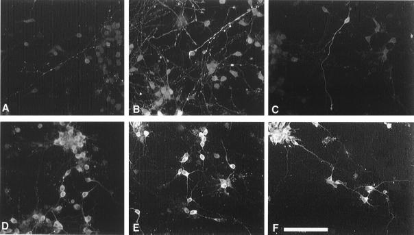
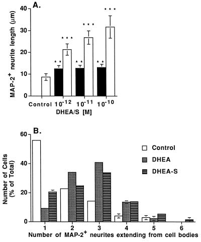
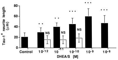
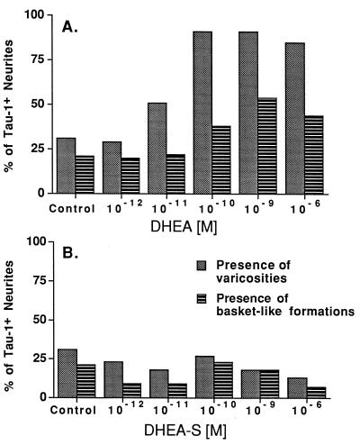
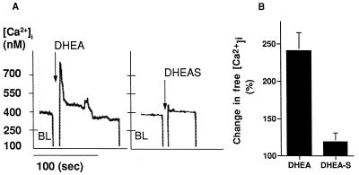
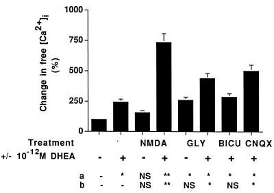
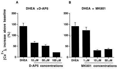
Dehydroepiandrosterone (DHEA) and its sulfate derivative (DHEAS) are the most abundant steroids produced by the human adrenal, but no receptors have been identified for these steroids, and no function for them has been established, other than as precursors for sex steroid synthesis. DHEA and DHEAS are found in brains from many species, and we have shown that enzymes crucial for their synthesis, especially P450c17 (17alpha-hydroxylase/c17,20 lyase), are expressed in a developmentally regulated, region-specific fashion in the developing rodent brain. One region of embryonic expression of P450c17, the neocortical subplate, has been postulated to play a role in guiding cortical projections to their appropriate targets. We therefore determined if products of P450c17 activity, DHEA and DHEAS, regulated the motility and/or growth of neocortical neurons. In primary cultures of mouse embryonic neocortical neurons, DHEA increased the length of neurites containing the axonal marker Tau-1, and the incidence of varicosities and basket-like process formations in a dose-dependent fashion. These effects could be seen at concentrations normally found in the brain. By contrast, DHEAS had no effect on Tau-1 axonal neurites but increased the length of neurites containing the dendritic marker microtubule-associated protein-2. DHEA rapidly increased free intracellular calcium via activation of N-methyl-D-aspartate (NMDA) receptors. These studies provide evidence of mechanisms by which DHEA and DHEAS exert biological actions, show that they have specific functions other than as sex steroid precursors, mediate their effects via non-classic steroid hormone receptors, and suggest that their developmentally regulated synthesis in vivo may play crucial and different roles in organizing the neocortex.


Figures

Figure 1
Figure 1
Immunostaining for Tau-1 (A–C) and MAP-2 (D–F) of E16.5 neocortical neurons cultured for 4 days. DHEA 10−12 M (B, E), DHEAS 10−12 M (C and F), or vehicle (0.1% dimethyl sulfoxide) (A and D) were added to the cultures for the last 20 h. DHEA promotes the abundance of Tau-1+ neurites (B). Vehicle or DHEAS has no effect on Tau-1+ neurites. Very few cell bodies were immunostained with the anti-Tau-1 antibody and no MAP-2+ dendrites were double-stained with the anti-Tau-1 antibody, suggesting that in our cultures, the cellular localization of Tau-1 is restricted to axons. However, the immunostaining with the anti-MAP-2 antibody was cytoplasmic and extended into dendrites. DHEA and DHEAS (E and F) increased MAP-2 staining in the cell bodies, but only DHEAS increased MAP-2+ neurite length (F). There are many faintly MAP-2+ collaterals and clustering of the neurons when cells are cultured with DHEAS. (Bar = 100 μm).
Figure 2
Figure 2
Dose dependent effect of DHEAS on MAP-2+ neurite outgrowth (A) and effect of steroids on the number of neurites extending from cell bodies (B). (A) Each column shows the mean length of MAP-2+ neurites for at least 100 labeled neurons/condition from three separate wells in one representative experiment. Open bars are DHEAS treatment and solid bars are DHEA treatment. Error bars are ±SD. Neurite length was quantitated by using the nih image 1.57 program. ∗∗∗, P < 0.0001, and ∗∗, P < 0.001 using a one-way ANOVA, with a Scheffe’s post hoc analysis. Three independent experiments gave similar results. (B) Histogram representing the percentage of cells with a specific number (–6) of MAP-2+ neurites was used to depict data from 10−11 M DHEA and 10−11 M DHEAS treatments. Experiments were performed at 10−12–10−10 M DHEA and DHEAS, and the original data were analyzed by one-way ANOVA, by using Scheffe’s post hoc analysis to determine individual differences at each concentration of steroid. ANOVA showed that DHEA and DHEAS treatments significantly increased the number of MAP-2+ neurites extending from cell bodies compared with untreated control cells. No significant differences were found among the different doses of DHEA and DHEAS, or between DHEA and DHEAS treatments. Open bars are control untreated cells, dark gray bars are DHEA treatment, and hatched bars are DHEAS treatment.
Figure 3
Figure 3
Dose dependent effect of DHEA on Tau-1+ neurite outgrowth. Each column shows the mean length of Tau-1+ neurites for at least 100 labeled neurons/condition from three separate wells in one representative experiment. Solid bars are DHEA treatment and open bars are DHEAS treatment. Error bars are ±SD. Neurite length was quantitated by using the nih image 1.57 program. Statistical differences were determined by using a one-way ANOVA with a post hoc Scheffe’s analysis; ∗∗∗, P < 0.0001; ∗∗, P < 0.001. Three independent experiments gave similar results.
Figure 4
Figure 4
Effect of DHEA (A) and DHEAS (B) on varicosities and basket-like formations on Tau-1+ neurites. The histograms express a single percentage of all neurons sampled. Gray bars are the percentage of Tau-1+ neurites bearing one or more varicosities; hatched bars are the percentage of Tau-1+ neurites making basket-like formations around cultured neurons. Data were analyzed by χ2 analysis. For DHEA treatment, χ2 = 131.4; P < 0.0001 for frequency of varicosities and χ2 = 30.3; P < 0.0001 for frequency of basket-like formations. The presence of varicosities and of basket-like formations is independent of each dose of DHEAS treatment (χ2 = 3.8; P < 0.567 for frequency of varicosities and χ2 = 4.7; P < 0.446 for frequency of basket-like formations).
Figure 5
Figure 5
DHEA increases [Ca2+]i measured by microfluorometry on a suspension of the cells preloaded with Indo-1. (A) shows a typical response of cells incubated with 10−12 M DHEA. Fluorescence was measured continuously for 3 min immediately following injection of the steroid. (B) Quantitation of peak [Ca2+]i was determined after calibration of the dye. There was a linear correlation between the values of the baseline and the values of the DHEA induced peak and plateau (peak: r2 = 0.879, P = 0.021; plateau: r2 = 0.977, P = 0.008), allowing us to correct peak and plateau values obtained in different experiments with respect to the baseline values. Therefore, we standardized the [Ca2+] for each experiment placing the baseline arbitrarily at 100% and presented the mean quantitative values (solid bars) as percent of variation of the peak value from the baseline. Error bars are ±SEM of the standardized [Ca2+] from at least three experiments, each performed in duplicate.
Figure 6
Figure 6
Effect of NMDA, glycine and receptor antagonists on the increase of [Ca2+]i mediated by DHEA. Each bar shows the percent mean [Ca2+]i increase over baseline. Error bars are ±SEM of six separate measurements. NMDA (10 μM), glycine (10 μM), or antagonists (10 μM) were added to the cell suspension before addition of DHEA. NMDA increased [Ca2+]i above baseline values, and DHEA synergistically potentiated this response; glycine increased [Ca2+]i, and DHEA increased this response. Bicuculline (BICU) and 6-cyano-7-nitroquinoxaline-2,3-dione (CNQX) did not block the response of the cells to DHEA. These results support the involvement of the NMDA receptor in the DHEA-mediated increase in [Ca2+]i. Statistical differences between (a) control and treatments and between (b) DHEA treatment and other treatments (shown in a table at the bottom of the figure), were determined by using a two-way ANOVA test and post hoc Scheffe’s analysis; ∗∗, P < 0.001; ∗, P < 0.05.
Figure 7
Figure 7
Dose-dependent effect of NMDA receptor antagonists D-AP5 (A) and MK801 (B) on the stimulation of [Ca2+]i mediated by DHEA. Each bar shows the percent mean [Ca2+]i change over the baseline. Error bars are ±SEM of at least three experiments.

Comment in

References

    1. Baulieu E E. Biol Cell. 1991;71:3–10. - PubMed
    1. Mellon S H. J Clin Endocrinol Metab. 1994;78:1003–1008. - PubMed
    1. Le Goascogne C, Robel P, Gouezou M, Sananes N, Baulieu E E, Waterman M. Science. 1987;237:1212–1215. - PubMed
    1. Mellon S H, Deschepper C F. Brain Res. 1993;629:283–292. - PubMed
    1. Dupont E, Simard J, Luu-The V, Labrie F, Pelletier G. Mol Cell Neurosci. 1994;5:119–123. - PubMed

Publication types

MeSH terms

LinkOut - more resources