. 1998 May 1;18(9):3138-46.
doi: 10.1523/JNEUROSCI.18-09-03138.1998.

G-Protein-coupled modulation of presynaptic calcium currents and transmitter release by a GABAB receptor

Affiliations

G-Protein-coupled modulation of presynaptic calcium currents and transmitter release by a GABAB receptor

T Takahashi et al. J Neurosci. .

Abstract

Presynaptic GABAB receptors play a regulatory role in central synaptic transmission. To elucidate their underlying mechanism of action, we have made whole-cell recordings of calcium and potassium currents from a giant presynaptic terminal, the calyx of Held, and EPSCs from its postsynaptic target in the medial nucleus of the trapezoid body of rat brainstem slices. The GABAB receptor agonist baclofen suppressed EPSCs and presynaptic calcium currents but had no effect on voltage-dependent potassium currents. The calcium current-EPSC relationship measured during baclofen application was similar to that observed on reducing [Ca2+]o, suggesting that the presynaptic inhibition generated by baclofen is caused largely by the suppression of presynaptic calcium influx. Presynaptic loading of the GDP analog guanosine-5'-O-(2-thiodiphosphate) (GDPbetaS) abolished the effect of baclofen on both presynaptic calcium currents and EPSCs. The nonhydrolyzable GTP analog guanosine 5'-O-(3-thiotriphosphate) (GTPgammaS) suppressed presynaptic calcium currents and occluded the effect of baclofen on presynaptic calcium currents and EPSCs. Photoactivation of GTPgammaS induced an inward rectifying potassium current at the calyx of Held, whereas baclofen had no such effect. We conclude that presynaptic GABAB receptors suppress transmitter release through G-protein-coupled inhibition of calcium currents.


Figures
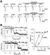
Fig. 1.
Fig. 1.
Inhibitory effects of GABAB receptor agonists on EPSCs. EPSCs were evoked by extracellular stimulation.A (top row), Reversible inhibition of EPSCs by baclofen (2 μm) and attenuation of the baclofen effect by CGP35348 (100 μm) in an MNTB neuron.Bottom row, Inhibitory effect of GABA (20 μm) and its attenuation by CGP35348 in another MNTB neuron. The magnitude of inhibition by CGP35348 on the effect of baclofen and GABA was 82.8 ± 2.3% (n = 3) and 60.7 ± 11% (n = 3), respectively.B, Dose-dependent suppression of EPSCs by baclofen and GABA. Cumulative dose-dependent effects of baclofen (top) and GABA (bottom) on the amplitude of EPSCs recorded from MNTB neurons. Sample records from individual MNTB neurons are shown in the inset. Calibration: 2 nA, 10 msec. The curves fitted to data points derived from the following equation: magnitude of inhibition (%) = maximal inhibition (%)/[1 + (IC50/agonist concentration)n]. For baclofen and GABA, maximal inhibition was 82.0 and 90.8%, IC50 was 0.77 and 9.97 μm, and Hill coefficient (n) was 0.90 and 1.21, respectively. Magnitude of EPSC suppression by 20 μm baclofen was 80.1 ± 2.6% at the cumulative-dose application (n = 4), which was not significantly different (p = 0.13; Student’s t test) from that at the single-dose application (72.5 ± 4.3%;n = 8).
Fig. 2.
Fig. 2.
Baclofen-induced suppression of EPSCs is blocked by GDPβS. Simultaneous presynaptic and postsynaptic recordings at the calyx–MNTB synapse. EPSCs were evoked by action potentials elicited by a depolarizing current pulse (2–10 msec) applied to a calyx through a whole-cell patch pipette. The postsynaptic holding potential was −70 mV. A, Reversible suppression of EPSCs by baclofen (20 μm). B, Blocking effect of GDPβS (tri-lithium salt, 3 mm) in the pipette on baclofen-induced suppression of EPSCs (a, b). A lower concentration of GDPβS (0.2 mm) did not prevent the effect of baclofen (data not shown). After the pipettes were retracted, a second paired recording was made from the same structures with a presynaptic pipette containing GTP (0.5 mm) instead of GDPβS. Baclofen clearly suppressed the EPSCs (c, d), which gradually recovered after washout (e). Complete recovery of EPSCs took 5–10 min (Fig. 7). When LiCl (9 mm) was included in the presynaptic pipette the baclofen effect was not attenuated (not shown). The amplitudes of EPSCs were normalized against the mean of the first seven (with GDPβS) or six (with GTP) data points before baclofen application in each experiment; the data point represents means and the error bars represent SEMs derived from paired recording experiments at three different synapses. Vertical calibration scales indicate 80 mV for presynaptic membrane potentials (A and B) and 1.25 nA (A) or 0.6 nA (B) for EPSCs. Scale bars, 10 msec.
Fig. 3.
Fig. 3.
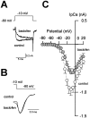
Suppression of presynaptic Ca2+currents by baclofen. The calyx was voltage-clamped at −80 mV, and IpCa was evoked by a 10 msec depolarizing pulse. In this experiment, [Ca2+]o was reduced to 1 mm to allow better voltage-clamp performance.A, IpCa induced in a calyx by a depolarizing voltage step to −10 mV in the absence and presence of baclofen (20 μm, superimposed). B, The tail currents are normalized at the peak and superimposed. C, Current–voltage relationships of IpCa before (open circles) and after (filled circles) baclofen application. Mean values ± SEMs obtained from six calyces are shown.
Fig. 4.
Fig. 4.
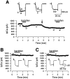
Block of baclofen-induced IpCa suppression by GTPγS or GDPβS. A, Occlusion of baclofen effect by GTPγS. IpCa was evoked in a calyx by a 20 msec depolarizing step from −70 mV to −13 mV. Baclofen (20 μm) suppressed IpCa, which recovered partially (a–c, superimposed). After a light flash given at an arrow for 2 sec, IpCa diminished gradually (c, d). A second application of baclofen after the flash had no effect on IpCa (d, e). Essentially the same result was obtained in two other calyces. B,C, Little effect of baclofen on IpCa (evoked by a 20 msec depolarizing pulse from −80 mV to −10 mV) was observed in the presence of GTPγS (200 μm, B) or GDPβS (3 mm, C) in the presynaptic pipette.B and C are from different calyces. A similar result was obtained in another calyx for each case.
Fig. 5.
Fig. 5.
Lack of baclofen effect on voltage-gated potassium currents. Inset, Outward potassium currents evoked by 20 mV depolarizing steps from the holding potential of −80 mV to +20 mV in the presence of TTX before (left) and after (middle) baclofen (20 μm) application. The superimposed traces before and after baclofen application overlapped almost completely (right). The amplitude of the potassium current was normalized against the value at 0 mV and mean ± SEMs of five calyces before (open circles) and after (filled triangles) baclofen application are plotted against membrane potential.
Fig. 6.
Fig. 6.
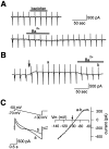
Effects of baclofen and GTPγS on presynaptic holding current and membrane conductance. A calyx was voltage-clamped at the holding potential of −70 mV, and a ramp command voltage from −50 to −130 mV (C, top left) was applied every 20 sec.A, Baclofen (20 μm) had no effect on the holding current or input conductance. Ba2+ (100 μm) caused a slight inward current associated with a decrease in conductance in the same calyx. B, In another calyx, photo-release of GTPγS by a UV flash (arrow) induced an outward current accompanied by an increase in input conductance. This current was suppressed by Ba2+(100 μm, b). Application of the light flash without loading caged compound had no effect on the holding current or membrane conductance. The outward current was not observed after GTPγS photolysis with the Cs+-based internal solution for IpCa recordings (Fig. 4A).C, Currents (a, b, bottom) corresponding to a command voltage (top) after photolysis of caged GTPγS compound before (a) and after (b) application of Ba2+.Right, Ba2+-sensitive current extracted as a difference current (a–b).Arrow indicates theoretical equilibrium potential for potassium ions calculated from the internal and external potassium activities. The difference current between before and after photolysis had a similar reversal potential, but inward rectification was less prominent (data not shown). Membrane potential was corrected for the liquid junction potential between the external and internal solution (+7.5 mV) for this current–voltage relationship. The data in this figure were low-pass-filtered at 100 Hz and sampled at 1 kHz.
Fig. 7.
Fig. 7.
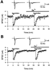
Effect of external Ba2+ and Cs+ on GABAB receptor-mediated presynaptic inhibition. EPSCs were evoked in MNTB principal cells by extracellular stimulation. A, Baclofen-induced suppression of EPSCs was similar before and after Ba2+ application (1 mm). EPSCs before, during baclofen application (20 μm), and after washout are superimposed on top in the absence (left) and presence (right) of Ba2+. Note the small polysynaptic EPSC component observed at the decay of monosynaptic EPSC. B, Baclofen suppressed EPSCs similarly in the absence and presence of external Cs+ (1 mm). EPSCs before and after baclofen application are superimposed on top in the absence (left) and presence (right) of Cs+. A andB are from different cells.
Fig. 8.
Fig. 8.
Comparison of IpCa–EPSC relationships during baclofen application and [Ca2+]oreduction. Paired recording from a calyx and its target cell.A, Effects of baclofen (20 μm; i, ii) and [Ca2+]o reduction (iii, iv) on IpCa (Pre) and EPSCs (Post). IpCa was evoked by 1 msec depolarizing command pulse from −70 mV to −10 mV. Records before and after baclofen application or [Ca2+]o reduction are superimposed on top row. B, Double logarithmic plot of IpCa–EPSC relation during baclofen application (filled circles with a dotted regression line) and [Ca2+]o reduction (open circles with a solid regression line). Data points above 90% in EPSC amplitude were excluded from these plots to minimize constrainment. The slope value was 2.29 for baclofen and 2.33 for [Ca2+]oreduction, respectively. Excluding the minimal point from each relationship had no significant effect on the slope values (2.15 and 2.09, respectively, for baclofen and [Ca2+]o reduction). Inset graph, The slope value of regression lines compared between [Ca2+]o reduction and baclofen application at seven synapses. No significant difference withp = 0.24 in paired t test. The mean slope value was 1.73 ± 0.17 for baclofen and 1.77 ± 0.17 for [Ca2+]o reduction, respectively.

References

    1. Allerton CA, Boden PR, Hill RG. Actions of the GABAB agonist, (−)-baclofen, on neurones in deep dorsal horn of the rat spinal cord in vitro. Br J Pharmacol. 1989;96:29–38. - PMC - PubMed
    1. Andrade R, Malenka RC, Nicoll RA. A G-protein couples serotonin and GABAB receptors to the same channels in hippocampus. Science. 1986;234:1261–1265. - PubMed
    1. Augustine GJ. Regulation of transmitter release at the squid giant synapse by presynaptic delayed rectifier potassium current. J Physiol (Lond) 1990;431:343–364. - PMC - PubMed
    1. Barnes-Davies M, Forsythe ID. Pre- and postsynaptic glutamate receptors at a giant excitatory synapse in rat auditory brainstem slices. J Physiol (Lond) 1995;488:387–406. - PMC - PubMed
    1. Borst JGG, Helmchen F, Sakmann B. Pre- and postsynaptic whole-cell recordings in the medial nucleus of the trapezoid body of the rat. J Physiol (Lond) 1995;489:825–840. - PMC - PubMed

Publication types

LinkOut - more resources