Comparative Study
. 1998 May 1;18(9):3171-9.
doi: 10.1523/JNEUROSCI.18-09-03171.1998.

Reversal of age-related alterations in synaptic plasticity by blockade of L-type Ca2+ channels

Affiliations
Comparative Study

Reversal of age-related alterations in synaptic plasticity by blockade of L-type Ca2+ channels

C M Norris et al. J Neurosci. .

Abstract

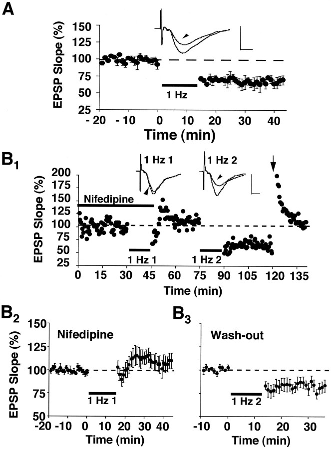
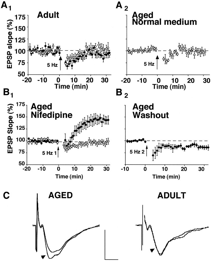
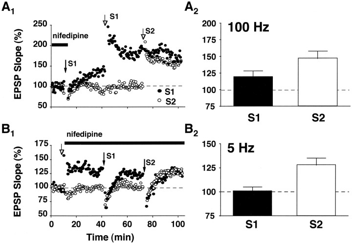
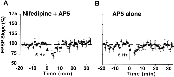
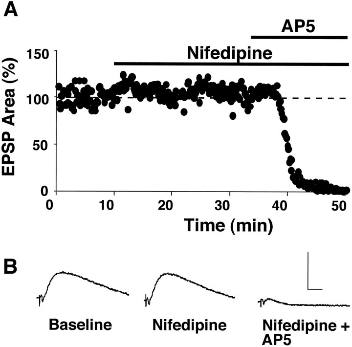
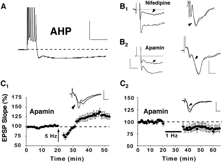
The role of L-type Ca2+ channels in the induction of synaptic plasticity in hippocampal slices of aged (22-24 months) and young adult (4-6 months) male Fischer 344 rats was investigated. Prolonged 1 Hz stimulation (900 pulses) of Schaffer collaterals, which normally depresses CA3/CA1 synaptic strength in aged rat slices, failed to induce long-term depression (LTD) during bath application of the L-channel antagonist nifedipine (10 microM). When 5 Hz stimulation (900 pulses) was used to modify synaptic strength, nifedipine facilitated synaptic enhancement in slices from aged, but not young, adult rats. This enhancement was pathway-specific, reversible, and impaired by the NMDA receptor (NMDAR) antagonist DL-2-amino-5-phosphonopentanoic acid (AP5). Induction of long-term potentiation (LTP) in aged rats, using 100 Hz stimulation, occluded subsequent synaptic enhancement by 5 Hz stimulation, suggesting that nifedipine-facilitated enhancement shares mechanisms in common with conventional LTP. Facilitation of synaptic enhancement by nifedipine likely was attributable to a reduction ( approximately 30%) in the Ca2+-dependent K+-mediated afterhyperpolarization (AHP), because the K+ channel blocker apamin (1 microM) similarly reduced the AHP and promoted synaptic enhancement by 5 Hz stimulation. In contrast, apamin did not block LTD induction using 1 Hz stimulation, suggesting that, in aged rats, the AHP does not influence LTD and LTP induction in a similar way. The results indicate that, during aging, L-channels can (1) facilitate LTD induction during low rates of synaptic activity and (2) impair LTP induction during higher levels of synaptic activation via an increase in the Ca2+-dependent AHP.


Figures

Fig. 1.
Fig. 1.
Nifedipine blocks the induction of LTD in aged rat slices. A, Plot of normalized field CA3/CA1 EPSP slopes (percentage of baseline ± SEM) obtained in stratum radiatum from three slices in response to stimulation of Schaffer collateral fibers (0.033 Hz). After an initial baseline period, 900 pulses of continuous 1 Hz stimulation (bar) induced a significant depression of the synaptic response. B1, An experiment conducted on an individual slice from an aged rat in which 1 Hz stimulation (1 Hz 1) was delivered in the presence of 10 μm nifedipine (thick bar) and did not depress the synaptic response. After the first 1 Hz train, nifedipine was washed from the recording medium, and a second episode of 1 Hz stimulation (1 Hz 2) was delivered in a drug-free medium. This second train of 1 Hz stimulation induced a marked depression that could be reversed to baseline by 100 Hz stimulus bursts (arrow). Insets for A andB1 display the averaged field EPSP waveforms from 10 successive responses collected immediately before and 30 min after (arrowheads) the delivery of 1 Hz stimulation. Calibration: 1 mV, 5 msec. B2,B3, Average data from aged rat slices (B2, n = 8;B3, n = 7) that were treated identically to the individual case inB1. Note that responses inB3 are normalized to the average of responses in B2 collected between 25 and 30 min after the termination of the first 1 Hz train.
Fig. 2.
Fig. 2.
Induction of synaptic modification by 5 Hz stimulation is a function of age, status of L-channel function, and synaptic strength. A1, Slices from adult rats displayed little change in the synaptic response after 5 Hz stimulation (arrow, 900 pulses), whether they were incubated in normal medium (open circles,n = 6) or in nifedipine-containing medium (filled circles, n = 7).A2, Similarly, 5 Hz stimulation delivered to aged rat slices (n = 5) bathed in normal perfusion medium also failed to modify synaptic strength.B1, In contrast, aged rat slices perfused with nifedipine exhibited a robust synaptic enhancement after 5 Hz stimulation (5 Hz 1) to the test pathway (filled circles, n = 16). Altered synaptic strength was not observed in the control pathways that did not receive 5 Hz stimulation (open circles, n = 14).B2, For four of the slices illustrated in B1, nifedipine was washed from the perfusion medium, and a second round of 5 Hz stimulation (5 Hz 2) was administered. In drug-free medium, depression of the enhanced response, attributable to 5 Hz stimulation, was obtained. Values plotted in B2 were normalized to the average of responses collected during the final 10 min of drug washout (i.e., the last 10 min of recording inB1). C, Shown are averaged waveforms (from 10 successive sweeps) collected from typical experiments in which 5 Hz stimulation was administered to slices bathed in nifedipine-containing medium. Arrowheads point to sweeps collected 25–30 min after the delivery of 5 Hz stimulation. Sweeps without arrowheads were collected during the 5 min immediately preceding the 5 Hz episode. Calibration: 1 mV, 5 msec.
Fig. 3.
Fig. 3.
Synaptic enhancement induced by 5 Hz stimulation during L-channel blockade is mechanistically similar to LTP induced by high-frequency stimulation. A1, Experiment conducted on an individual aged rat slice in which synaptic enhancement was first induced in one population of synapses (S1, filled arrow), using 5 Hz stimulation in the presence of nifedipine. After the 5 Hz episode, nifedipine was washed from the recording medium, and two bursts of 100 Hz stimulation were delivered to S1 (open arrow, S1). Approximately 30 min later, a second round of 100 Hz stimulation was delivered to naive synapses in a second input (open arrow, S2).A2, Average data from three aged rat slices in which induction of synaptic enhancement by 5 Hz stimulation preceded the induction of LTP by 100 Hz stimulation. Note that, although 100 Hz stimulation produced further potentiation in S1, the percentage of increase in the synaptic response was much less than the increase observed at naive synapses (i.e., S2) after 100 Hz stimulation. B1, For an individual aged rat slice, LTP was induced in one pathway (open arrow, S1), using 100 Hz stimulation. After induction of LTP, nifedipine was washed into the recording medium, and a round of 5 Hz stimulation was applied to the potentiated synapses (closed arrow, S1). At 30 min after the first round of 5 Hz stimulation, a second 5 Hz train was applied to nonpotentiated synapses (S2) in the continued presence of nifedipine.B2, Average data from five aged rat slices in which induction of LTP by 100 Hz stimulation preceded the induction of synaptic enhancement by 5 Hz stimulation. Note that, after the induction of LTP in S1, 5 Hz stimulation in the presence of nifedipine fails to induce synaptic enhancement. However, 5 Hz stimulation applied to nonpotentiated synapses in S2 produces robust enhancement. Error bars indicate SEM.
Fig. 4.
Fig. 4.
Synaptic enhancement induced by 5 Hz stimulation is sensitive to AP5. A, Aged rat slices (n = 5) that were coincubated with nifedipine and AP5 (100 μm) did not exhibit synaptic enhancement after a 5 Hz stimulus train. B, Similarly, 5 Hz stimulation failed to increase synaptic strength when aged rat slices (n = 4) were bathed in AP5 alone.
Fig. 5.
Fig. 5.
Nifedipine does not alter basal NMDAR-mediated responses. A, Plot of intracellular EPSPs (area above baseline) from an individual aged rat slice in response to stimulation of Schaffer collateral fibers (0.1 Hz). In this experiment the slice was incubated in DNQX (20 μm), picrotoxin (10 μm), and low extracellular Mg2+ (0.5 mm) to isolate the NMDAR component of CA3/CA1 synaptic transmission. After an initial 10 min baseline, nifedipine was applied to the slice and did not alter the EPSP. Subsequent application of AP5 (100 μm) dramatically reduced the EPSP, demonstrating successful isolation of the NMDAR component of synaptic transmission. B, Average intracellular EPSP waveforms generated from 10 successive responses collected during the initial baseline, 30 min after application of nifedipine, and 15 min after the addition of AP5. Calibration: 5 mV, 10 msec.
Fig. 6.
Fig. 6.
Nifedipine enhances synaptic strength via a reduction in the AHP. A, An example of an AHP recorded intracellularly from a CA1 pyramidal cell in an aged rat after a train of seven action potentials, elicited by a 100 msec pulse of depolarizing current. Calibration: 20 mV, 200 msec.B1, Illustrations of the AHP (cell held at −65 mV) after a burst of seven action potentials (left) and the field EPSP (right) before and after (arrowheads) application of nifedipine to the recording medium. B2, Illustrations of the AHP (cell held at −71 mV) after a burst of seven action potentials (left) and the field EPSP (right) before and after (arrowheads) application of the K+ channel blocker apamin (1 μm) to the recording medium. These data show that nifedipine and apamin reduce the AHP to a similar extent. However, neither drug substantially alters the EPSP slope. Waveforms inB are averages of five consecutive responses collected before and after drug wash-in. Also, in the left panels, note that action potentials were truncated to better illustrate the AHPs. Calibration: for AHPs, 2.5 mV, 200 msec; for EPSPs, 0.5 mV, 5 msec. C1, Like nifedipine, application of apamin to aged rat slices (n = 7) facilitated the induction of synaptic enhancement attributable to 5 Hz stimulation. C2, In contrast to nifedipine, apamin did not prevent the induction of LTD after 1 Hz stimulation (n = 7). Insets forC display the averaged field EPSP waveforms from 10 successive responses collected immediately before and 30 min after (arrowhead) the delivery of pattern stimulation. Calibration: 1 mV, 5 msec.

References

    1. Barnes CA, McNaughton BL. Spatial memory and hippocampal synaptic plasticity in senescent and middle-aged rats. In: Stein D, editor. The psychobiology of aging: problems and perspectives. Elsevier; Amsterdam: 1980. pp. 253–272.
    1. Barnes CA, Rao G, Foster TC, McNaughton BL. Region-specific age effects on AMPA sensitivity: electrophysiological evidence for loss of synaptic contacts in hippocampal field CA1. Hippocampus. 1992;2:457–468. - PubMed
    1. Barnes CA, Rao G, McNaughton BL. Functional integrity of NMDA-dependent LTP induction mechanisms across the lifespan of F-344 rats. Learn Memory. 1996;3:124–137. - PubMed
    1. Bear MF. Mechanism for a sliding synaptic modification threshold. Neuron. 1995;15:1–4. - PubMed
    1. Bear MF, Abraham WC. Long-term depression in hippocampus. Annu Rev Neurosci. 1996;19:437–462. - PubMed

Publication types