. 1998 May 1;18(9):3213-23.
doi: 10.1523/JNEUROSCI.18-09-03213.1998.

Stable complexes involving acetylcholinesterase and amyloid-beta peptide change the biochemical properties of the enzyme and increase the neurotoxicity of Alzheimer's fibrils

Affiliations

Stable complexes involving acetylcholinesterase and amyloid-beta peptide change the biochemical properties of the enzyme and increase the neurotoxicity of Alzheimer's fibrils

A Alvarez et al. J Neurosci. .

Abstract

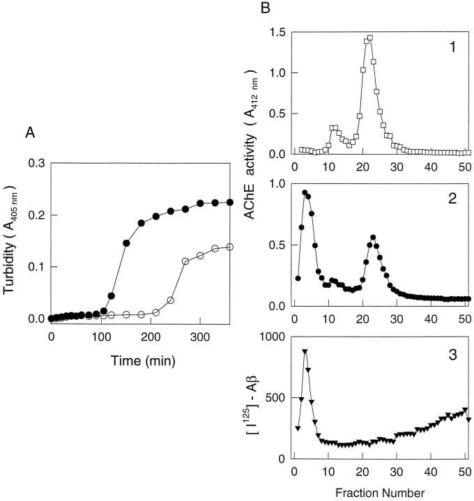
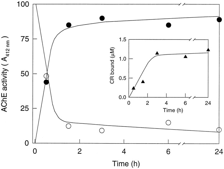
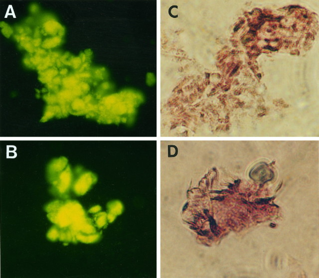
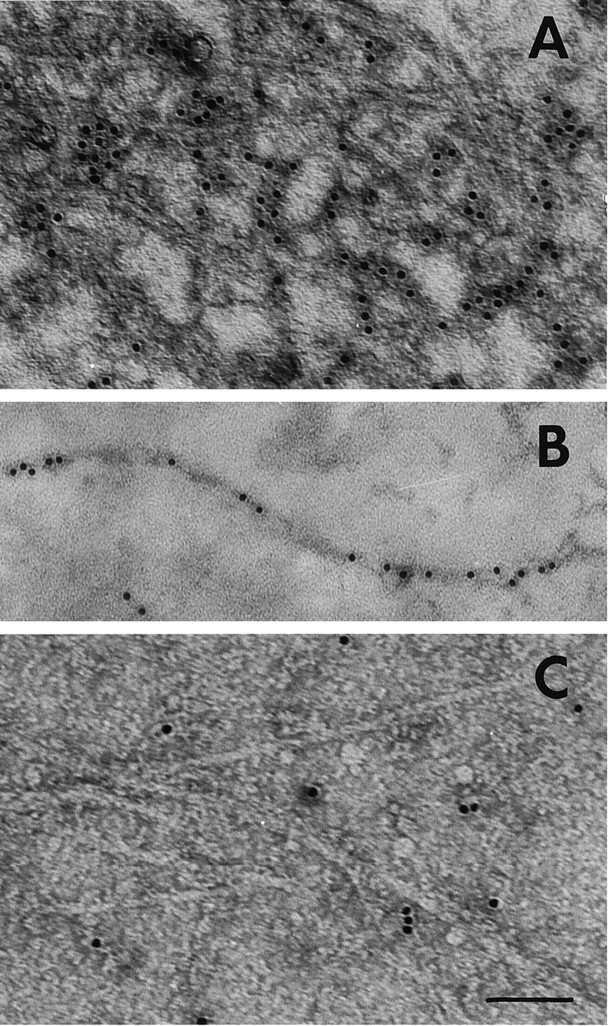
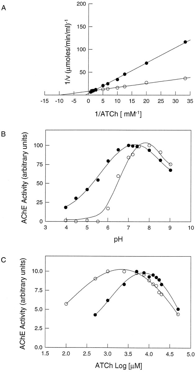
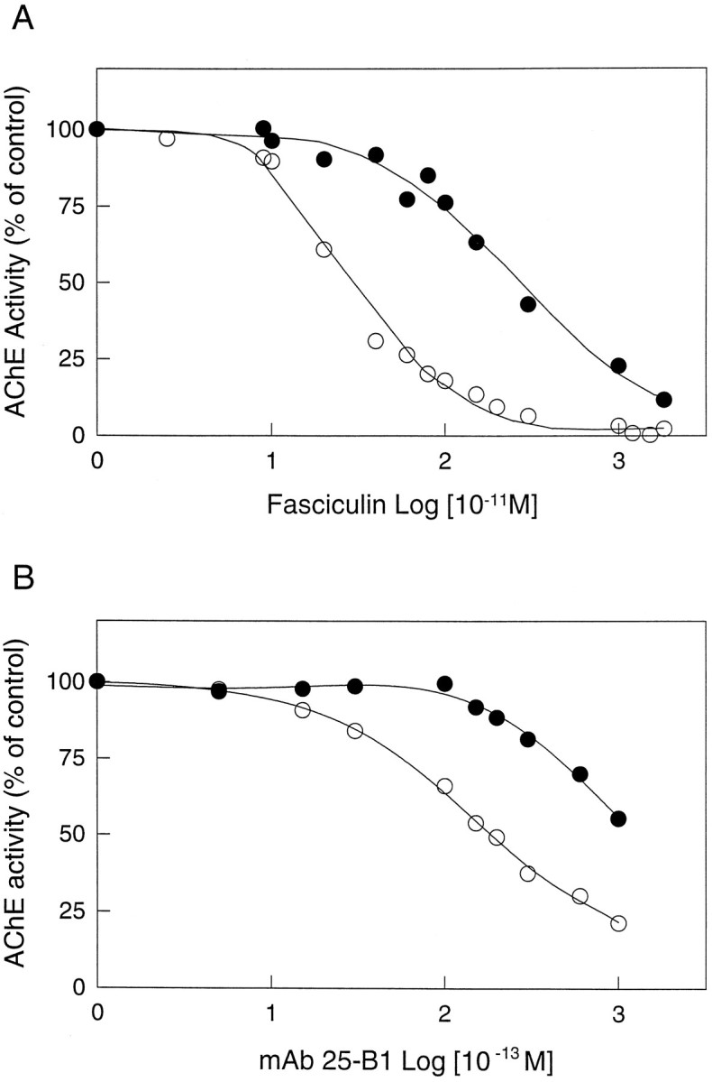
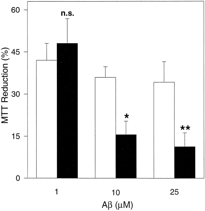
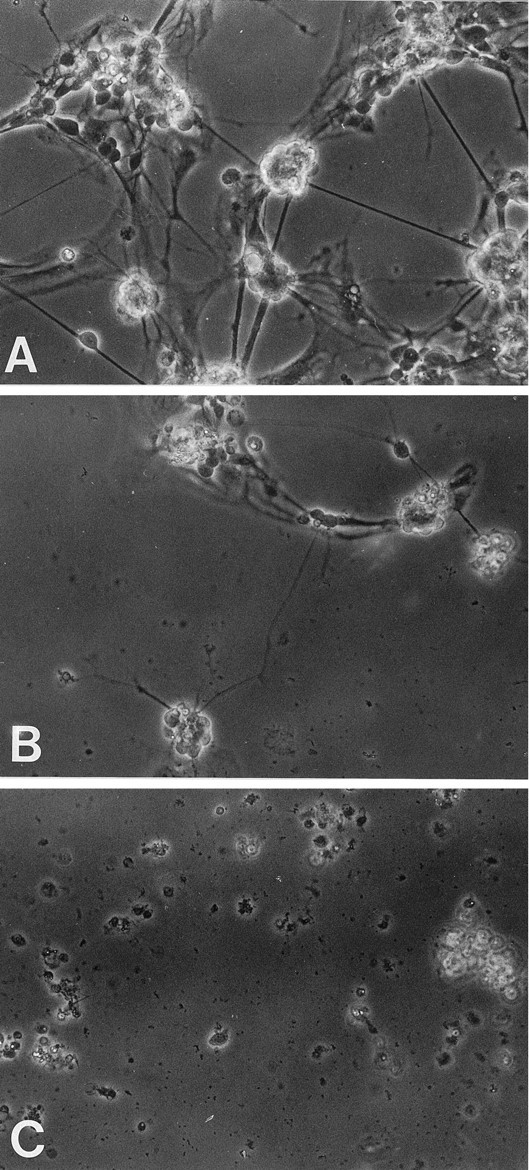
Brain acetylcholinesterase (AChE) forms stable complexes with amyloid-beta peptide (Abeta) during its assembly into filaments, in agreement with its colocalization with the Abeta deposits of Alzheimer's brain. The association of the enzyme with nascent Abeta aggregates occurs as early as after 30 min of incubation. Analysis of the catalytic activity of the AChE incorporated into these complexes shows an anomalous behavior reminiscent of the AChE associated with senile plaques, which includes a resistance to low pH, high substrate concentrations, and lower sensitivity to AChE inhibitors. Furthermore, the toxicity of the AChE-amyloid complexes is higher than that of the Abeta aggregates alone. Thus, in addition to its possible role as a heterogeneous nucleator during amyloid formation, AChE, by forming such stable complexes, may increase the neurotoxicity of Abeta fibrils and thus may determine the selective neuronal loss observed in Alzheimer's brain.


Figures

Fig. 1.
Fig. 1.
The AChE is associated with Aβ amyloid fibrils.A, Aβ peptide aggregation assay. Aβ peptide (97 μm in PBS, pH 7.4) was incubated in a kinetic stirred assay, with (•) or without (○) AChE (0.1 μm). Aβ aggregation was followed by turbidity at 405 nm. B, Sedimentation velocity analysis. Native brain AChE (10.7 S) as control and 125I-Aβ amyloid aggregates produced in a stirred kinetic assay with AChE were submitted to velocity sedimentation analysis in 5–20% sucrose gradients containing a 70% sucrose cushion at the bottom of the centrifuge tube. A Combi-Sorval ultracentrifuge was used. Fractions were collected from the bottom of the gradient as described previously (Inestrosa et al., 1994). AChE activity and radioactivity in the corresponding fractions were determined.1, Control G4 AChE; 2, AChE–Aβ aggregates; 3, 125I-Aβ amyloid aggregates.
Fig. 2.
Fig. 2.
Temporal dependence of AChE–Aβ fibril complex formation. Turbidimetric stirred assays of Aβ with AChE were incubated for 90 min. Then aliquots of 40 μl were taken at different incubation times, diluted in 400 μl of PBS, and centrifuged, and AChE activity was determined in the fibril pellet (•) and the supernatant fraction (○) by the assay of Ellman et al. (1961). The amyloid character of the aggregates formed in the presence of AChE was determined by Congo red assays (inset) in the pellet fraction.
Fig. 3.
Fig. 3.
Thioflavine-T fluorescence and AChE histochemical staining of the AChE–Aβ complexes. A, B, Amyloid aggregates formed in a kinetic stirred assay were washed three times with PBS and then incubated for 4 min in a solution containing an excess of thioflavine-T (5 mg/ml). Samples were examined under an Axioplan fluorescence microscope (D-7082; Zeiss, Oberkochen, Germany) at 400× magnification. C, D, Amyloid aggregates from a kinetic stirred assay were washed three times with PBS, after which the fibrils were stained for AChE with the histochemical staining reaction of Karnovsky and Roots (1964).
Fig. 4.
Fig. 4.
Immunogold labeling of the AChE–Aβ complexes.A, AChE–Aβ complexes were labeled with a polyclonal antibody raised against AChE, followed by an anti-IgG antibody conjugated to 10 nm colloidal gold particles, and were visualized by negative staining. B, Select example of an amyloid fiber labeled with anti-AChE conjugated to gold particles. C, Control sample of high molecular mass Aβ aggregates without AChE that were treated with the same specific antibody as above. In each case, 10 μl aliquots taken from an Aβ peptide kinetic stirred aggregation assay, performed either in the presence or absence of AChE, were adsorbed onto 300-mesh Formvar-coated grids, negative-stained with 2% uranyl acetate, and viewed for fibrils with a Philips electron microscope. Scale bar: A, C, 90 nm;B, 80 nm.
Fig. 5.
Fig. 5.
Biochemical characterization of the AChE activity associated with amyloid-β fibrils. AChE–Aβ complexes were washed exhaustively with PBS using four cycles of centrifugation and resuspension to remove noncomplexed AChE, and then amyloid-associated AChE activity was studied (•). Soluble native brain AChE was used as control (○). A, Lineweaver–Burk (double-reciprocal) plots. AChE activity, free and associated with amyloid-β fibrils, was measured over a range of substrate concentrations. B, pH dependence. Optimal pH for AChE and AChE–Aβ complex activity was determined over a pH range of 4.0–9.0 in phosphate buffer.C, Activity of AChE as a function of substrate concentration. The rate of hydrolysis is plotted as a log function of acetylthiocholine substrate concentration. The bell-shaped curves show that both free AChE and the AChE–Aβ complexes are inhibited by excess substrate. However, the AChE–Aβ complexes require higher acetylthiocholine concentrations for optimal activity than the free enzyme.
Fig. 6.
Fig. 6.
Inhibition of the AChE–Aβ complex by fasciculin and the monoclonal antibody 25-B1 (mAb 25-B1), two peripheral anionic binding site ligands. The activity is plotted as a function of fasciculin (A) and mAb 25-B1 (B) concentrations. Free AChE (○) and AChE–Aβ complexes (•) are both inhibited by fasciculin and mAb 25-B1. However, the AChE–Aβ complexes require higher concentrations of both compounds to reach the same level of inhibition as free AChE. The IC50 values for free AChE were 249 ± 15 pm for fasciculin and 19.0 ± 0.5 pm for mAb 25–B1. The corresponding values for the AChE–Aβ complexes were 2746 ± 28 pm for fasciculin and >100 pmfor mAb 25–B1. The IC50 values represent the mean ± SD of three identical samples run in separate experiments.
Fig. 7.
Fig. 7.
AChE–Aβ complexes are more toxic than Aβ fibrils alone in PC12 cells. Rat pheochromocytoma PC12 cells were treated with Aβ amyloid fibrils as a control (white bars) or with AChE–Aβ amyloid fibrils (black bars) for 48 hr at different Aβ concentrations. The cell viability after treatment was measured by the MTT reduction assay. Significantly different from control: *p < 0.05; **p < 0.001 by nonpaired Student’st test; n.s., not significant.
Fig. 8.
Fig. 8.
AChE–Aβ complexes are more toxic than Aβ fibrils alone in primary cultures of retina cells. Six-day chick retina cell cultures were incubated for 24 hr in a medium supplemented with PBS as a control (A), β-amyloid fibrils (2.5 μm) (B), and AChE–Aβ complexes (2.5 μm for the Aβ peptide) (C). The retina cell cultures were observed by phase contrast microscopy using 40× magnification under an Olympus inverted microscope. Representative photographs are shown.

References

    1. Abraham CR, Selkoe DJ, Potter H. Immunochemical identification of the serine protease inhibitor α1-antichymotrypsin in the brain amyloid deposits of the Alzheimer’s disease. Cell. 1988;52:487–501. - PubMed
    1. Alvarez A, Bronfman F, Pérez CA, Vicente M, Garrido J, Inestrosa NC. Acetylcholinesterase, a senile plaque component, affects the fibrillogenesis of amyloid-β-peptides. Neurosci Lett. 1995;201:49–52. - PubMed
    1. Alvarez A, Opazo C, Alarcon R, Garrido J, Inestrosa NC. Acetylcholinesterase promotes the aggregation of amyloid-β-peptide fragments by forming a complex with the growing fibrils. J Mol Biol. 1997;272:348–361. - PubMed
    1. Arispe N, Pollard HB, Rojas E. Giant multilevel cation channels formed by Alzheimer disease amyloid β-protein [AβP-(1–40)] in bilayer membrane. Proc Natl Acad Sci USA. 1993;90:10573–10577. - PMC - PubMed
    1. Barcikowska M, Wisniewski HM, Bancher C, Grundke-Iqbal I. About the presence of paired helical filaments in dystrophic neurites participating in the plaque formation. Acta Neuropathol (Berl) 1989;78:225–231. - PubMed

Publication types

LinkOut - more resources