. 1998 May 1;18(9):3273-81.
doi: 10.1523/JNEUROSCI.18-09-03273.1998.

Competitive signaling between TrkA and p75 nerve growth factor receptors determines cell survival

Affiliations

Competitive signaling between TrkA and p75 nerve growth factor receptors determines cell survival

S O Yoon et al. J Neurosci. .

Abstract

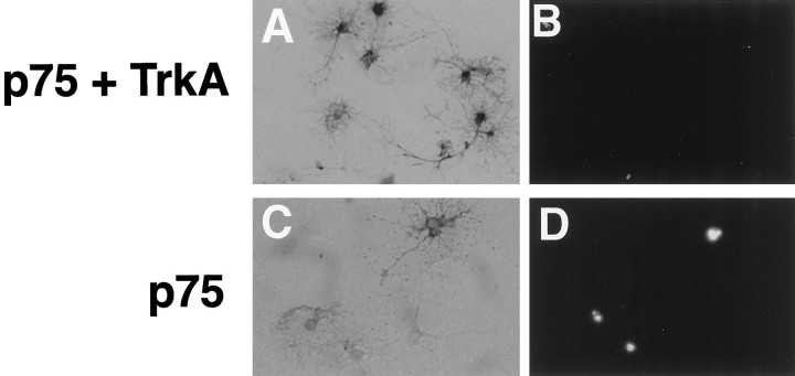
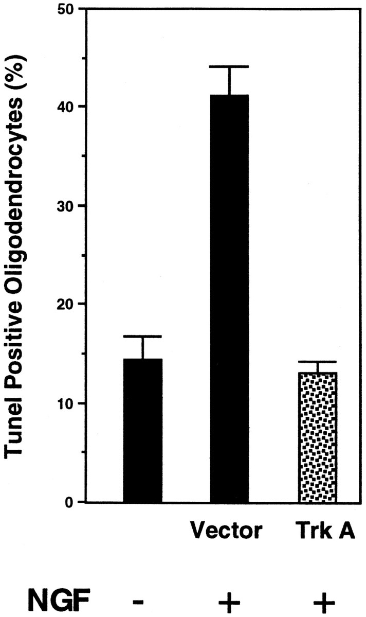
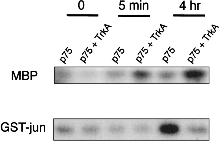
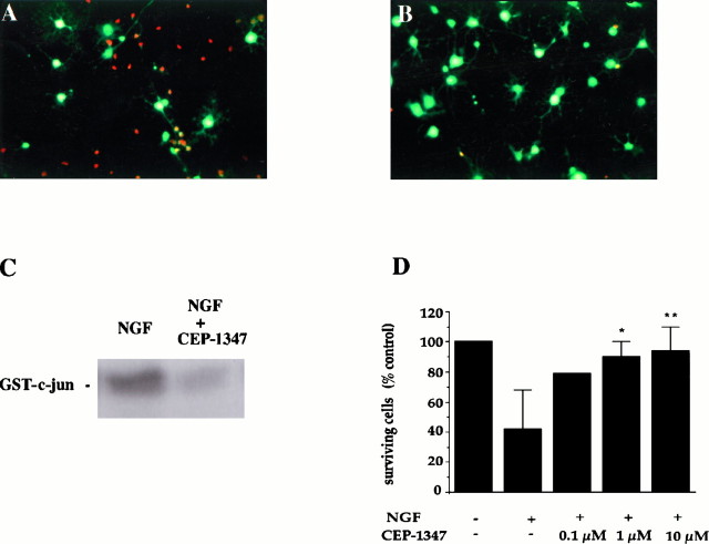
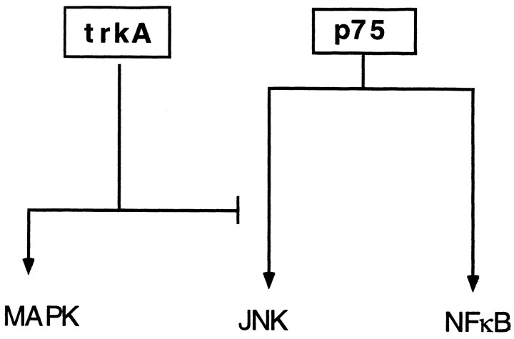
In addition to its role as a survival factor, nerve growth factor (NGF) has been implicated in initiating apoptosis in restricted cell types both during development and after terminal cell differentiation. NGF binds to the TrkA tyrosine kinase and the p75 neurotrophin receptor, a member of the tumor necrosis factor cytokine family. To understand the mechanisms underlying survival versus death decisions, the TrkA receptor was introduced into oligodendrocyte cell cultures that undergo apoptosis in a p75-dependent manner. Here we report that activation of the TrkA NGF receptor in oligodendrocytes negates cell death by the p75 receptor. TrkA-mediated rescue from apoptosis correlated with mitogen-activated protein kinase activation. Concurrently, activation of TrkA in oligodendrocytes resulted in suppression of c-jun kinase activity initiated by p75, whereas induction of NFkappaB activity by p75 was unaffected. These results indicate that TrkA-mediated rescue involves not only activation of survival signals but also simultaneous suppression of a death signal by p75. The selective interplay between tyrosine kinase and cytokine receptors provides a novel mechanism that achieves alternative cellular responses by merging signals from different ligand-receptor systems.


Figures

Fig. 1.
Fig. 1.
Expression of human TrkA receptors in oligodendrocytes after retroviral infection. A, Diagram of the recombinant TrkA retrovirus. The human TrkA cDNA was subcloned upstream of an IRES-linked alkaline phosphatase gene.B, Ligand-dependent activation and expression of TrkA in oligodendrocytes. Differentiated oligodendrocytes infected with TrkA (TrkA) or null (Con) retroviruses were either treated or untreated with 100 ng/ml NGF for 5 min. Tyrosine phosphorylation of TrkA receptors was detected after immunoprecipitation with an anti-Trk (203) antibody and Western blot with anti-phosphotyrosine antibodies 4G10 and PY20 (PY). The level of receptor expression under each condition was determined by reprobing the same blot with the anti-Trk 203 antibody. C, TrkA expression is induced by mitogenic stimuli. The expression of TrkA mRNA was monitored by reverse transcription-PCR using RNA isolated from oligodendrocyte progenitors and mature oligodendrocytes cultured in different conditions. A 129 bp fragment was amplified for rat trkA mRNA. Progenitors were maintained for 4 d either in DMEM supplemented with 20% B104 conditioned medium (B104) or with 2 ng/ml basic FGF and PDGF (PDGF/FGF) before RNA harvesting. Oligodendrocytes were differentiated either from progenitors directly placed in serum-free defined medium (Differentiation Medium) or from progenitors expanded in PDGF and bFGF (−PDGF/FGF). TrkA mRNA is induced by mitogenic stimuli and persists after removal of the growth factors.
Fig. 2.
Fig. 2.
Expression of p75 in oligodendrocytes infected with the trkA retrovirus. Differentiated oligodendrocytes infected with trkA retrovirus were stained for alkaline phosphatase (a) and p75 receptors (b) using the 9651 anti-p75 polyclonal antibody (Huber and Chao, 1995a). Cultures were allowed to grow in oligodendrocyte differentiation media for 7 d before staining. Cell shown was TUNEL-negative. Magnification, 400×.
Fig. 3.
Fig. 3.
Oligodendrocytes infected with TrkA or null virus. Cells infected with either the TrkA virus (A,B) or control null virus (C,D) were identified by staining for alkaline phosphatase (A, C). B, D, TUNEL staining of the same field of cells. Magnification, 200×.
Fig. 4.
Fig. 4.
TrkA prevents p75-mediated apoptosis. Quantification of TrkA-mediated rescue was performed after the TUNEL assay, as described in Materials and Methods. The number of TUNEL-positive cells in similar untreated cultures is indicated. In total, 500–600 cells were counted from four separate experiments.
Fig. 5.
Fig. 5.
Activation of TrkA shifts the balance between MAP kinases and JNK. Control differentiated cultures infected with the control virus (p75+) and cultures infected with the TrkA virus (p75 +TrkA) were treated or left untreated with 100 ng/ml NGF for 5 min or 4 hr. Lysates were prepared and subjected to immunoprecipitation/kinase assays, using MBP as a substrate for MAP kinase activity and GST-jun as a substrate for JNK activity. The experiments were repeated three times with similar results.
Fig. 6.
Fig. 6.
Inhibition of JNK activity prevents NGF-induced cell death in oligodendrocytes. Double immunofluorescence of cultures treated for 4 hr with 100 ng of NGF (A) or 100 ng of NGF plus 1 μm CEP-1347 (B) and then stained live with ethidium and calcein AM. Rednuclei indicate dying cells, and green fluorescent cells reflect surviving oligodendrocytes. C, c-Jun kinase assay on cell lysates obtained from cultures treated with 100 ng/ml NGF and 100 ng/ml NGF plus 1 μm CEP-1347 for 4 hr. The experiment was performed in duplicate. D, Quantitation of surviving cells (number of green fluorescent cells) after 4–8 hr treatment with NGF (100 ng/ml) or NGF (100 ng/ml) plus CEP-1347 at increasing concentrations. The results represent the mean ± SEM of the cells from 6 to 10 determinations (except for controls and 100 nm CEP-1347 that was performed in duplicate). *p = 0.002; **p = 0.001.
Fig. 7.
Fig. 7.
Top, Nuclear translocation of RelA/p65 after NGF treatment in p75+ cultures. Cultures were either left untreated (A,B) or treated with 100 ng/ml NGF for 1 hr (C, D) and then fixed in ethanol-formaldehyde. The expression of RelA/p65 was assessed by indirect immunofluorescence using monoclonal antibodies against the activated form of the RelA subunit and streptoavidin-Cy3.A, C, Phase-contrast photomicrographs.B, D, Immunostaining with anti-RelA antibody. Bottom, NFκB activation in oligodendrocytes. TrkA expression does not affect NFκB activation by p75. DNA binding activity of NFκB in control and TrkA-expressing oligodendroglial cultures is shown. Lysates were prepared from cultures and assessed for electrophoretic gel mobility shift using a 32P-labeled oligonucleotide with a κ light chain enhancer sequence.
Fig. 8.
Fig. 8.
Model for competitive signaling between TrkA and p75 receptors in oligodendrocytes. NGF-binding to p75 can induce NFκB and JNK activation, whereas TrkA activates MAP kinase phosphorylation. When the two receptors are expressed together, TrkA blocks the p75-mediated signaling leading to JNK activation. On the other hand, NFκB activation by p75 is left unaffected.

References

    1. Althaus HH, Kloppner S, Schmidt-Schultz T, Schwartz P. Nerve growth factor induces proliferation and enhances fiber regeneration in oligodendrocytes isolated from adult pig brain. Neurosci Lett. 1992;135:219–223. - PubMed
    1. Baeuerle PA, Baltimore D. NF-κB: ten years after. Cell. 1996;87:13–20. - PubMed
    1. Barbacid M. The trk family of neurotrophin receptors. J Neurobiol. 1994;25:1386–1403. - PubMed
    1. Barker PA, Shooter EM. Disruption of NGF binding to the low-affinity neurotrophin receptor p75 reduces NGF binding to trkA on PC12 cells. Neuron. 1994;13:203–215. - PubMed
    1. Barrett GL, Bartlett PF. The p75 receptor mediates survival or death depending on the stage of sensory neuron development. Proc Natl Acad Sci USA. 1994;91:6501–6505. - PMC - PubMed

Publication types