. 1998 May 1;18(9):3314-26.
doi: 10.1523/JNEUROSCI.18-09-03314.1998.

Development of bone morphogenetic protein receptors in the nervous system and possible roles in regulating trkC expression

Affiliations

Development of bone morphogenetic protein receptors in the nervous system and possible roles in regulating trkC expression

D Zhang et al. J Neurosci. .

Abstract

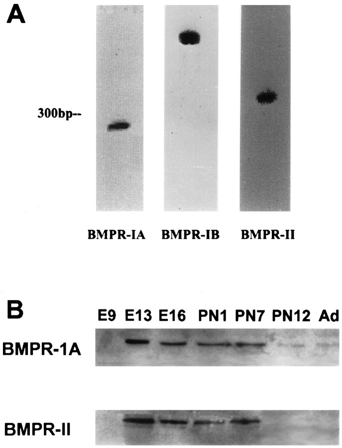
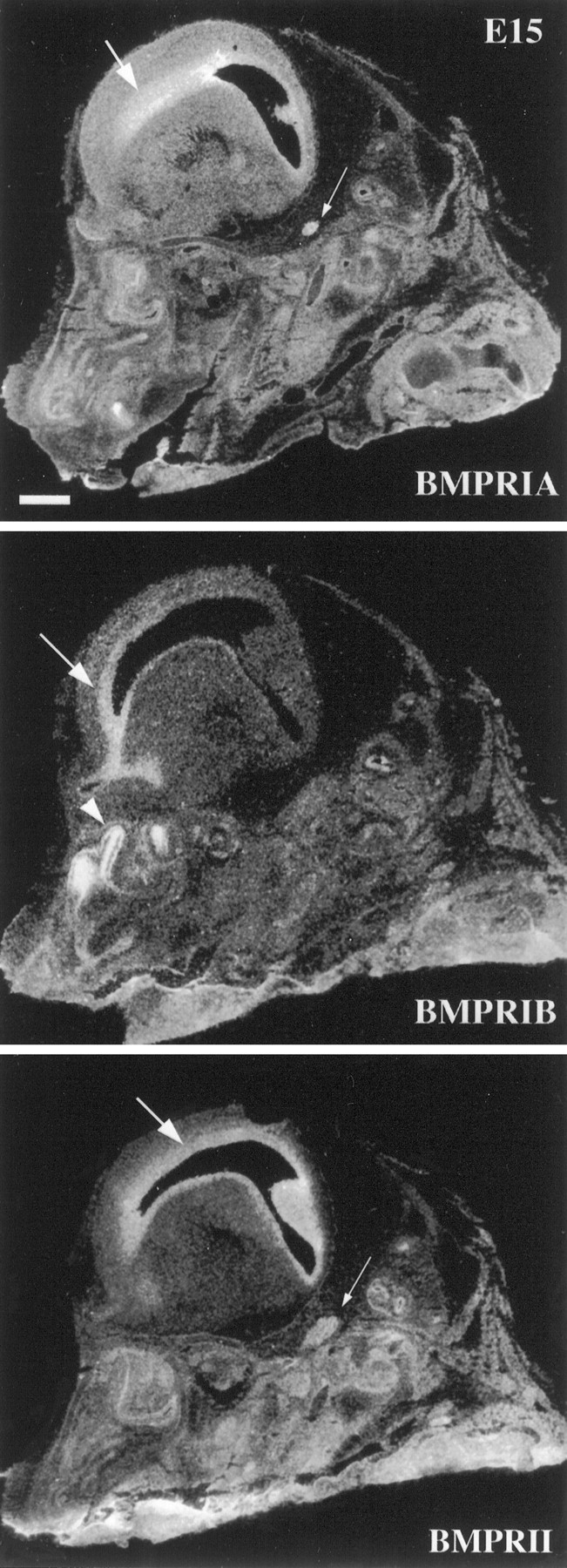
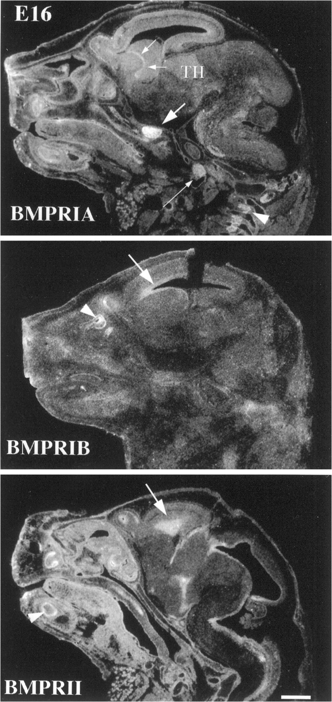
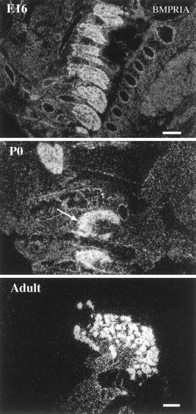
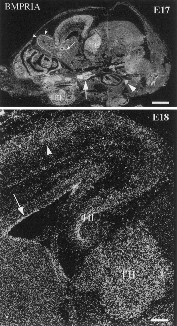
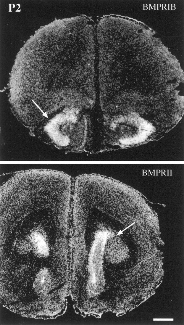
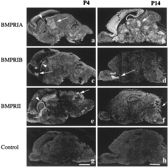
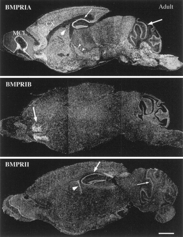
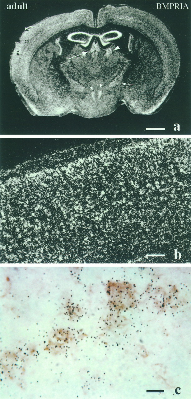
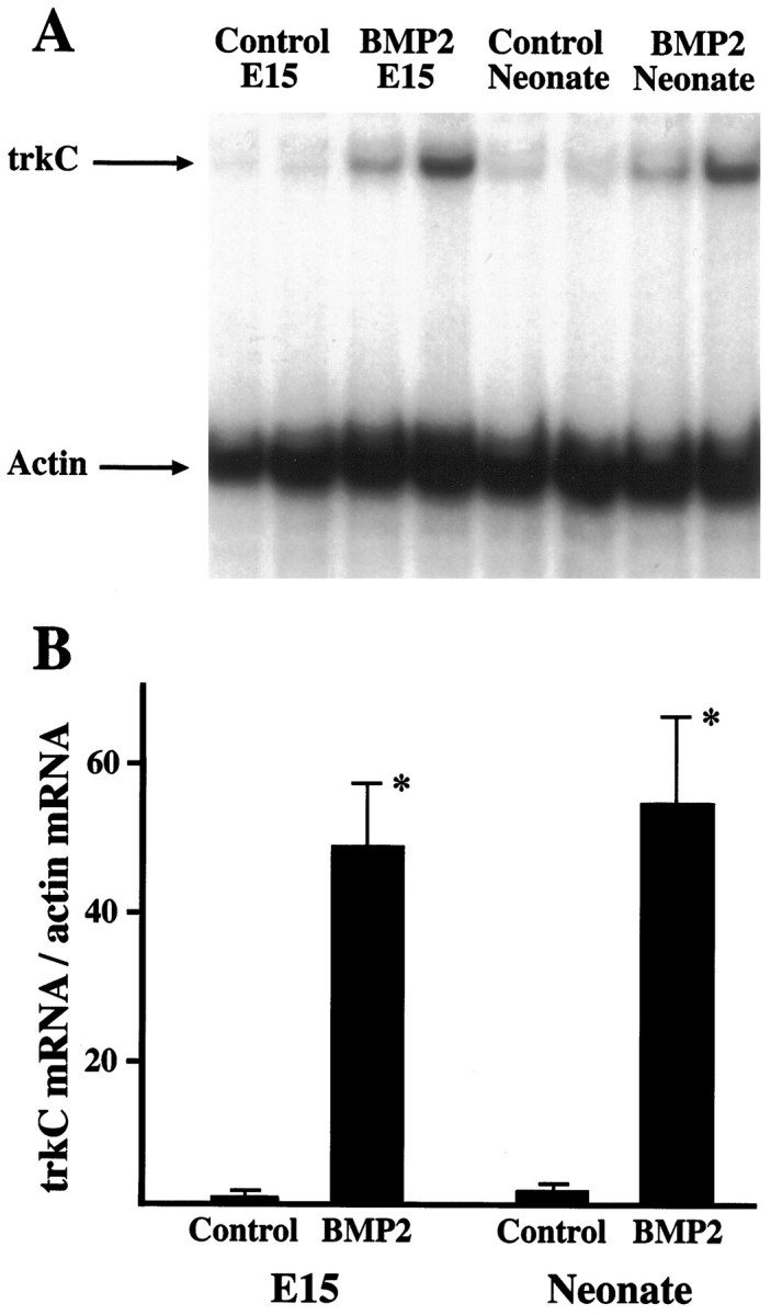
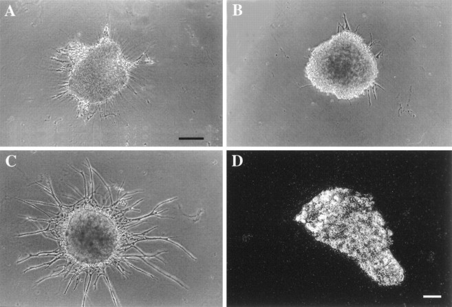
Characterization of bone morphogenetic protein receptor (BMPR) expression during development is necessary for understanding the role of these factors during neural maturation. In this study, in situ hybridization analyses demonstrate that BMP-specific type I (BMPR-IA and BMPR-IB) and type II (BMPR-II) receptor mRNAs are expressed at significant levels in multiple regions of the CNS, cranial ganglia, and peripheral sensory and autonomic ganglia during the embryonic and neonatal periods. All three BMP receptor subunits are expressed within periventricular generative zones. BMPR-IA is more abundant than the other receptor subtypes, with widespread expression in the brain, cranial ganglia, and peripheral ganglia. By contrast, BMPR-IB mRNA displays significant expression within more restricted regions, including the anterior olfactory nuclei. BMPR-II mRNA exhibits peak expression within the cerebellar Purkinje cell layer and the hippocampus, as well as within cranial ganglia. The distribution of BMP receptors within large neurons in adult dorsal root ganglia suggested a possible role in regulating expression of the neurotrophin receptor trkC. This hypothesis was tested in explant cultures of embryonic day 15 (E15) and postnatal day 1 (P1) sympathetic superior cervical ganglia (SCG). Treatment of the E15 or the P1 SCG with BMP-2 induced expression of trkC mRNA and responsiveness of sympathetic neurons to NT3 as measured by neurite outgrowth. The pattern of expression of BMP receptors in embryonic brain suggests several potentially novel areas for further developmental analysis and supports numerous recent studies that indicate that BMPs have a broad range of cellular functions during neural development and in adult life.


Figures

Fig. 1.
Fig. 1.
A, Expression of BMPR-IA, BMPR-IB, and BMPR-II mRNAs in neonatal mouse whole brain samples as detected by RNase protection assays.B, Western blot analyses of BMPR-IA and BMPR-II proteins in embryonic (E9, E13, E16), postnatal (P1, P7, P12), and adult (Ad) whole brains.
Fig. 2.
Fig. 2.
Expression of BMP receptor subunit mRNAs as detected by in situ hybridization and radioautography in sagittal sections of E15 mouse head. BMPR-IA mRNA is expressed by the VZ (arrow) and the trigeminal ganglion (small arrow) in the nervous system. The expression ofBMPR-IB mRNA is restricted to the frontal region of the VZ (arrow) and the olfactory epithelium (arrowhead). BMPR-II mRNA is also expressed in the VZ (arrow) and the trigeminal ganglion (small arrow). Scale bar, 1 mm.
Fig. 3.
Fig. 3.
Expression of BMP receptor subunit mRNAs in the E16 mouse head. BMPR-IA mRNA is expressed at high levels in the trigeminal ganglion (arrow), nodose ganglion (small arrow), and DRG (arrowhead). It is also expressed by the striatum and the contiguous periventricular generative zone (small arrows). Relatively weak and diffuse expression is found in the thalamus (TH). Expression of BMPR-IB mRNA is limited to the frontal region of the periventricular generative zone (arrow) and to the olfactory epithelium (arrowhead).BMPR-II mRNA is also expressed by the periventricular generative zone (arrow). Scale bar, 1 mm.
Fig. 4.
Fig. 4.
Expression of BMPR-IA mRNA in DRG during development. BMPR-IA mRNA is expressed homogeneously in DRG (cervical region) in the E16 mouse embryo. The abundant expression persists in P0 DRG (arrow, cervical region) and in Adult DRG (shown at higher magnification). Notice that in Adult, BMPR-IA mRNA is expressed primarily by large neuronal cells. Scale bars: E16 and P0, 300 μm;Adult, 100 μm.
Fig. 5.
Fig. 5.
Expression of BMPR-IA mRNA in theE17 and E18 mouse head. BMPR-IA mRNA is expressed abundantly by the trigeminal ganglion (arrow) and nodose ganglion (arrowhead) at E17. It is also expressed homogeneously in the thalamus, mitral cell layer (small arrowheads), striatal generative zone (small arrows), cortical plate, and the primordium of the teeth. Note that in the high-power image of E18mouse brain, BMPR-IA mRNA is expressed in the thalamus, hippocampus, VZ/SVZ (arrow), and cortical plate (arrowhead). TH, Thalamus;HI, hippocampus; PT, primordium of the teeth. Scale bars: E17, 1 mm; E18, 200 μm.
Fig. 6.
Fig. 6.
Expression of BMPR mRNAs within frontal cortical regions of the P2 mouse brain. BMPR-IB mRNA expression is limited to the anterior olfactory nuclei (arrow), whereas BMPR-II is expressed at high levels within the region of the SVZ (arrow). Scale bar, 500 μm.
Fig. 7.
Fig. 7.
Expression of BMP receptor mRNAs at postnatal days 4 and 14. BMPR-IA mRNA is widely distributed in theP4 (a) and P14(b) mouse brains. Notice that the expression pattern within the thalamus changes from a homogeneous pattern atP4 (arrow) to expression within specific nuclei at P14 (arrow). The expression ofBMPR-IB is restricted to the anterior olfactory nuclei at P4 (c) and P14(d). The expression of BMPR-II is seen within the hippocampus, cerebellum (arrow), and mitral cell layer of the olfactory bulb at P4(e) and P14(f). Notice that all three receptors are specifically expressed within the ependymal layer of the SVZ atP4 (a, c, e; arrowhead), but not at older stages, such as P14 (b, d, f; arrowhead). BMPR-II is expressed within the cerebellum in the postnatal period at higher levels than BMPR-IA. g, h, Controlsections hybridized with BMPR-IA sense probe showing background labeling. Scale bars: a, c, e, g, 2 mm; b, d, f, h, 2 mm.
Fig. 8.
Fig. 8.
The expression of BMP receptor subunit mRNAs as detected by in situ hybridization and radioautography in sagittal sections of adult mouse brain. BMPR-IA mRNA is widely expressed throughout the brain. Intense labeling is seen within the cerebral cortex (CX), hippocampus (small arrow), Purkinje cell layer of the cerebellum (arrow), brainstem, thalamic nuclei (small arrow heads), mitral cell layer of olfactory bulb (MCL), and choroid plexus of the lateral ventricle (arrowhead). The expression of BMPR-IBmRNA is restricted to the anterior olfactory nuclei (arrow). BMPR-II mRNA is expressed at modest levels within the hippocampus (arrow), choroid plexus (arrowhead), Purkinje cell layer of the cerebellum (small arrow), and the mitral cell layer of the olfactory bulb. Scale bar, 2 mm.
Fig. 9.
Fig. 9.
Expression of BMPR-IA mRNA in the adult mouse brain. a, Coronal section of adult brain through the region of the thalamus. Notice that BMPR-IA mRNA is expressed in medial dorsal and paraventricular thalamic nuclei (arrow), lateral dorsal nuclei (arrowhead), zona incerta (small arrows), and dorsal medial hypothalamic nucleus (small arrowhead). b, c, Cerebral cortical regions at high magnification. Notice inc that BMPR-IA mRNA is colocalized with immunostaining for neurofilament, indicative of expression in neurons. Scale bars:a, 1 mm; b, 200 μm;c, 100 μm.
Fig. 10.
Fig. 10.
Regulation of trkC expression by BMP-2.A, Explants of E15 and of neonatal superior cervical ganglia were cultured in the absence or presence of BMP-2 (10 ng/ml) for 48 hr and were examined by nuclease protection assay for levels of trkC mRNA. Levels of actin mRNA were simultaneously determined to assure equivalent loading of RNA in each lane. Note thatBMP-2 treatment increased levels of trkCmRNA in both E15 and neonatal explants.B, Levels of trkC mRNA in the nuclease protection assays were quantitated densitometrically and normalized to levels of actin mRNA in each sample. The level inE15 control ganglia was assigned a value of 1, and other samples are normalized to this control value. *Differs from respective control value at p < 0.01; n = 4.
Fig. 11.
Fig. 11.
Effects of BMP-2 treatment on neurite extension by sympathetic ganglia. E15 sympathetic ganglia were cultured in the absence of added factors or with NT3 (20 ng/ml; A), BMP-2 (20 ng/ml; B), or NT3 and BMP-2 (C). Like untreated ganglia (data not shown), neurite outgrowth was minimal with NT3 alone or BMP-2 alone but was much more extensive after combined treatment with NT3 and BMP-2.D, In situ hybridization analysis of an adult SCG for BMPR-1A mRNA. Note that the mRNA is still expressed abundantly even in the adult. Scale bars in A–C, 130 μm.

Similar articles

Cited by

References

    1. Arkell R, Beddington RSP. BMP-7 influences pattern and growth of the developing hindbrain of mouse embryos. Development. 1997;124:1–12. - PubMed
    1. Birren SJ, Anderson DJ. A V-myc-immortalized sympathoadrenal progenitor cell line in which neuronal differentiation is initiated by FGF but not NGF. Neuron. 1990;4:189–201. - PubMed
    1. Coughlin MD, Collins MB (1985) Nerve growth factor-independent development of embryonic mouse sympathetic neurons in dissociated cell culture Dev Biol 110:392–401. - PubMed
    1. D’Alessandro JS, Vetz-Aldape J, Wang EA. Bone morphogenetic proteins induce differentiation in astrocyte lineage cells. Growth Factors. 1994;11:53–69. - PubMed
    1. Dale L, Howes G, Price BM, Smith JC. Bone morphogenetic protein 4: a ventralizing factor in early Xenopus development. Development. 1992;115:573–585. - PubMed

Publication types

MeSH terms

LinkOut - more resources