. 1998 May 1;18(9):3327-35.
doi: 10.1523/JNEUROSCI.18-09-03327.1998.

Development of the mouse inner ear and origin of its sensory organs

Affiliations

Development of the mouse inner ear and origin of its sensory organs

H Morsli et al. J Neurosci. .

Abstract

The molecular mechanisms dictating the morphogenesis and differentiation of the mammalian inner ear are largely unknown. To better elucidate the normal development of this organ, two approaches were taken. First, the membranous labyrinths of mouse inner ears ranging from 10.25 to 17 d postcoitum (dpc) were filled with paint to reveal their gross development. Particular attention was focused on the developing utricle, saccule, and cochlea. Second, we used bone morphogenetic protein 4 (BMP4) and lunatic fringe (Fng) as molecular markers to identify the origin of the sensory structures. Our data showed that BMP4 was an early marker for the superior, lateral, and posterior cristae, whereas Fng served as an early marker for the macula utriculi, macula sacculi, and the sensory portion of the cochlea. The posterior crista was the first organ to appear at 11.5 dpc and was followed by the superior crista, the lateral crista, and the macula utriculi at 12 dpc. The macula sacculi and the cochlea were present at 12 dpc but became distinguishable from each other by 13 dpc. Based on the gene expression patterns, the anterior and lateral cristae may share a common origin. Similarly, three sensory organs, the macula utriculi, macula sacculi, and cochlea, seem to arise from a single region of the otocyst.


Figures

Fig. 1.
Fig. 1.
Lateral view of paint-filled membranous labyrinths ranging from 10.75 to 17 dpc. The scale bar and orientation shown inA also apply to B and C. The scale bar and orientation in D apply toE and F. G shows all inner ears at the same magnification, with arrows illustrating the growth of the proximal part of the cochlea from 13 to 17 dpc. Theinsets at the bottom left ofCF are ventral views of the cochlea. The orientation shown in inset of C also applies to insets in DF.Arrows point to the proximal part of the cochlea;arrowheads point to the distal part of the cochlea.Asterisks point to areas of reabsorption in the central regions of the developing superior and posterior canals.cc, Common crus; co, cochlea;csd, cochleosaccular duct; ed, endolymphatic duct; es, endolymphatic sac;hp, horizontal canal plate; la, lateral ampulla; lsc, lateral semicircular canal;pa, posterior ampulla; psc, posterior semicircular canal; s, saccule; sa, superior ampulla; ssc, superior semicircular canal;u, utricle; usd, utriculosaccular duct;vpl, vertical canal plate. Scale bars, 100 μm.
Fig. 2.
Fig. 2.
Top. Gene expression patterns ofBMP4 and Fng in developing mouse inner ear from 9 to 10.25 dpc by whole-mount in situhybridization. At 9 dpc, BMP4 expression was detected in the posterior margin of the otic cup (A,arrow), whereas Fng transcripts were in the most ventral part of the otic cup (D,arrow). At 9.5 dpc, BMP4 was diffusely expressed in the posterior part of the otocyst (B,arrow). Fng transcripts were localized to the most anteroventral part of the otocyst (E,arrow). At 10.25 dpc, BMP4 was expressed in two distinct areas, a posterior focus and an anterior streak (C, arrow, arrowhead, respectively). Fng transcripts were restricted to the most anteroventral quadrant of the otocyst (F,arrow). Orientation: A, anterior;D, dorsal. Scale bar, 100 μm.
Fig. 4.
Fig. 4.
Gene expression patterns of BMP4and Fng in the developing inner ear at 12 dpc. All panels are horizontal sections such that the anterior part of the embryo is toward the top. The level of each section is represented in the ear diagram at the bottom right.F and F′ are 12 μm adjacent sections.BMP4 was expressed in four distinct areas. The anterior streak of BMP4 signal at 11.5 dpc had now split into the presumptive superior and lateral cristae (A,sc, lc), and BMP4 was still expressed in the posterior crista (B,pc). The lco of BMP4became more elaborate, originating in the lateral part of the inner ear and expanding into the greater curvature of the cochlea (D, E, F,lco). Fng was expressed in two distinct areas. The most dorsal area was the presumptive macula utriculi (C, mu). The most ventral area was in the cochlea, where the signal originated in the medial part of the basal turn expanding into the lesser curvature of the cochlea (E′, F′, mco).BMP4 and Fng were coexpressed in a small area at the tip of the cochlea (F, F′,brackets). NT-3 gene expression overlapped with that of Fng in the cochlea (F"). lc, Lateral crista;lco, lateral cochlear hybridization signal;mco, medial cochlear hybridization signal;mu, macula utriculi; pc, posterior crista; sc, superior crista. Orientation:A, anterior; L, lateral. Scale Bar, 100 μm.
Fig. 6.
Fig. 6.
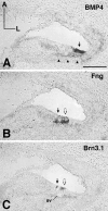
Gene expression of BMP4,Fng, and Brn-3.1 in developing cochlea at P1. AC are 12 μm adjacent sections.BMP4 transcripts were localized to specialized cells lateral to the outer hair cells: Hensen’s and/or Claudius’ cells (A, arrow). BMP4 was also expressed in the mesenchyme surrounding the cochlea (A,arrowheads). Fng transcripts were restricted to the supporting cells underneath the inner and outer hair cells (B, arrow, open arrow, respectively). Brn-3.1 was expressed in the inner and outer hair cells (C, arrow,open arrow, respectively). sv, Spiral vessel. Orientation: A, anterior; L, lateral. Scale Bar, 100 μm.

References

    1. Bast TH, Anson BJ. The temporal bone and the ear, pp 30–105. Thomas; Springfield, IL: 1949. The otic labyrinth.
    1. Bissonnette JP, Fekete DM. Standard atlas of the gross anatomy of the developing inner ear of the chicken. J Comp Neurol. 1996;174:1–11. - PubMed
    1. Cohen B, Bashirullah A, Dagnino L, Campbell C, Fisher WW, Leow CC, Whiting E, Ryan D, Zinyk D, Boulianne G, Hui CC, Gallie B, Phillips RA, Lipshitz HD, Egan SE. Fringe boundaries coincide with Notch-dependent patterning centres in mammals and alter Notch-dependent development in Drosophila. Nat Genet. 1997;16:283–288. - PubMed
    1. Cohen GM, Cotanche DA. Development of the sensory receptors and their innervation in the chick cochlea. In: Romand AL, editor. Development of auditory and vestibular systems, Vol 2. Elsevier; New York: 1992. pp. 101–138.
    1. Erkman L, McEvilly RJ, Luo L, Ryan AK, Hooshmand F, O’Connell SM, Keithley EM, Rapaport DH, Ryan AF, Rosenfeld MG. Role of transcription factors Brn-3.1 and Brn-3.2 in auditory and visual system development. Nature. 1996;381:603–606. - PubMed

Publication types

LinkOut - more resources