. 1998 May 1;18(9):3386-403.
doi: 10.1523/JNEUROSCI.18-09-03386.1998.

GABAergic cells are the major postsynaptic targets of mossy fibers in the rat hippocampus

Affiliations

GABAergic cells are the major postsynaptic targets of mossy fibers in the rat hippocampus

L Acsády et al. J Neurosci. .

Abstract

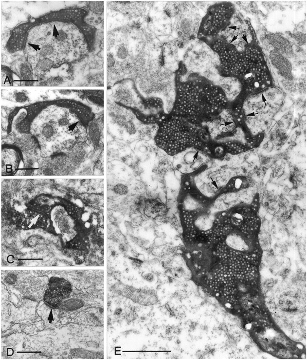
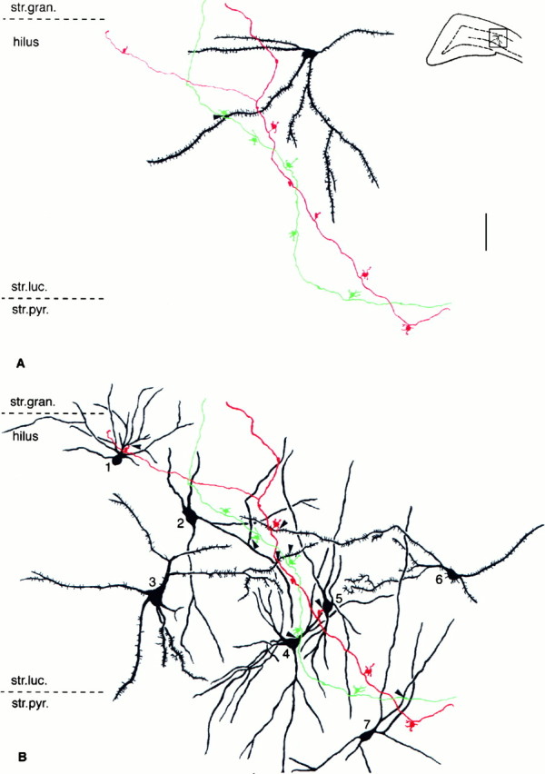
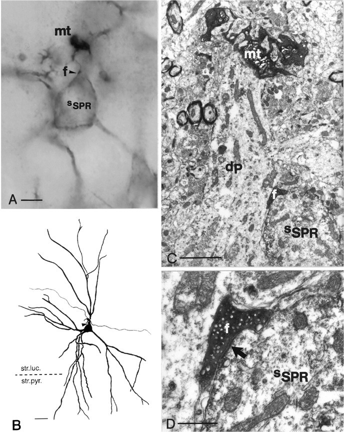
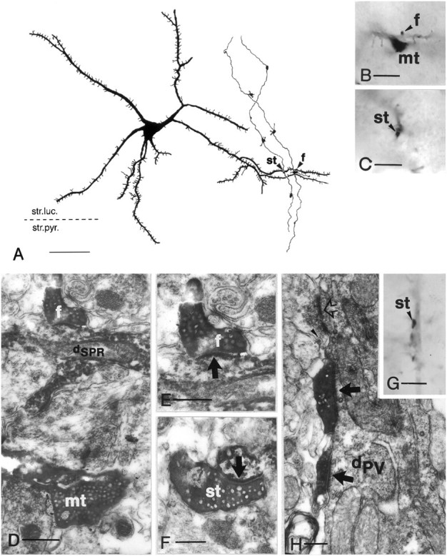
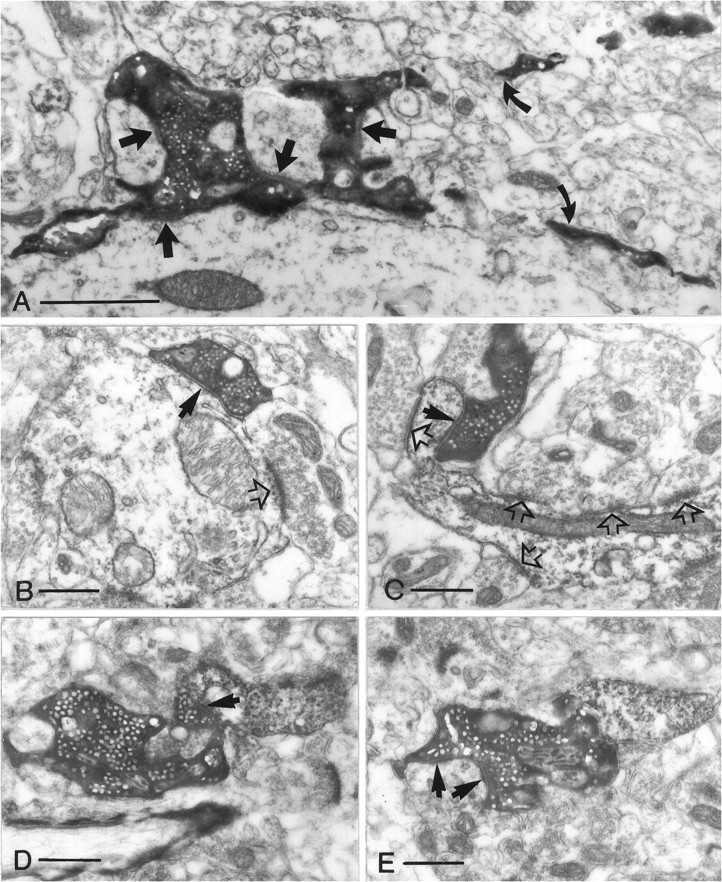
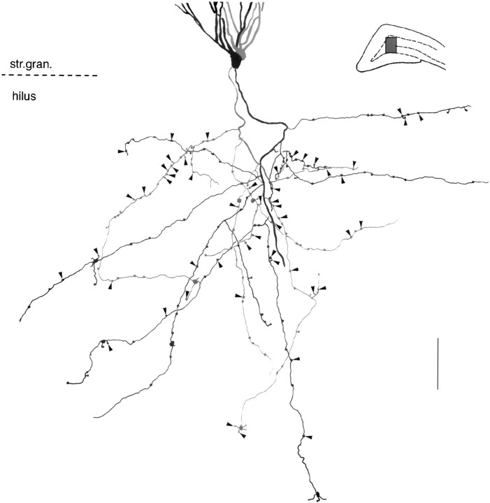
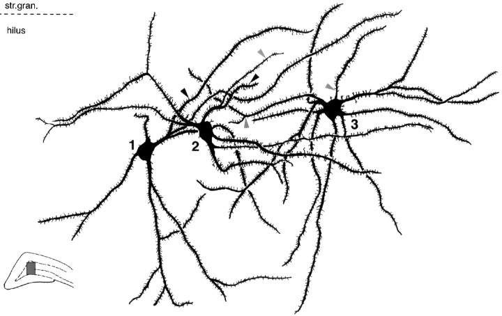
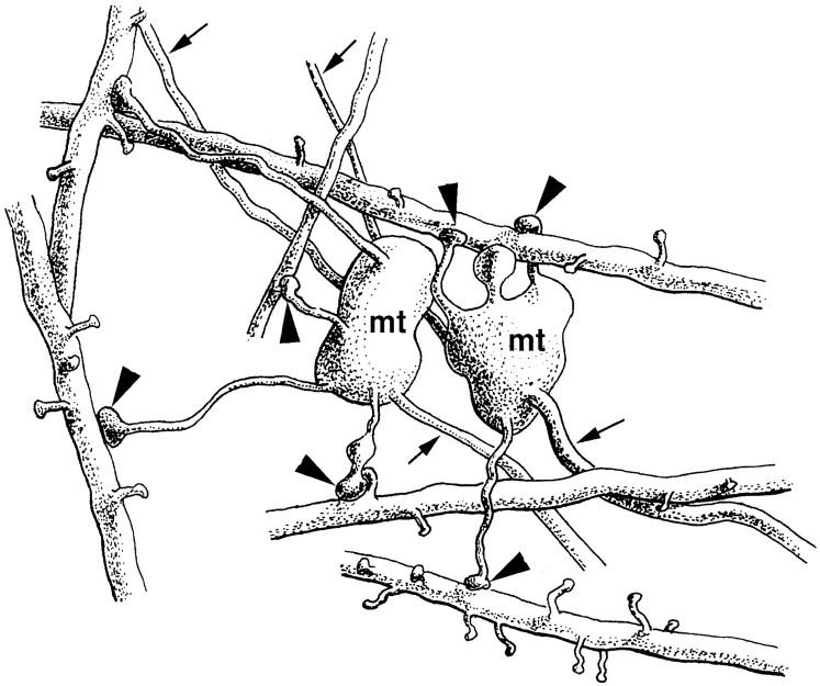
Dentate granule cells communicate with their postsynaptic targets by three distinct terminal types. These include the large mossy terminals, filopodial extensions of the mossy terminals, and smaller en passant synaptic varicosities. We examined the postsynaptic targets of mossy fibers by combining in vivo intracellular labeling of granule cells, immunocytochemistry, and electron microscopy. Single granule cells formed large, complex "mossy" synapses on 11-15 CA3 pyramidal cells and 7-12 hilar mossy cells. In contrast, GABAergic interneurons, identified with immunostaining for substance P-receptor, parvalbumin, and mGluR1a-receptor, were selectively innervated by very thin (filopodial) extensions of the mossy terminals and by small en passant boutons in both the hilar and CA3 regions. These terminals formed single, often perforated, asymmetric synapses on the cell bodies, dendrites, and spines of GABAergic interneurons. The number of filopodial extensions and small terminals was 10 times larger than the number of mossy terminals. These findings show that in contrast to cortical pyramidal neurons, (1) granule cells developed distinct types of terminals to affect interneurons and pyramidal cells and (2) they innervated more inhibitory than excitatory cells. These findings may explain the physiological observations that increased activity of granule cells suppresses the overall excitability of the CA3 recurrent system and may form the structural basis of the target-dependent regulation of glutamate release in the mossy fiber system.


Figures

Fig. 1.
Fig. 1.
Granule cell filled with biocytin in vivo. The cell was first developed for biocytin and photographed, and the image was fused with the cresyl violet-stained image of the same section. Arrow indicates mossy fiber;white arrowheads indicate local collaterals in the hilar region. m, Molecular layer; g, granule cell layer; h, hilus; CA3, pyramidal layer of CA3c. Top inset, Current–voltage responses of the granule cell to hyperpolarizing (−0.8, −0.6, and −0.4 nA) and depolarizing (0.7 and 0.8 nA) current steps. Bottom inset, High magnification of a mossy terminal (arrow) in the hilar region. Double arrowhead indicates filopodial extension; white arrowheads indicate small en passant and drumstick-like boutons. Scale bar, 10 μm.
Fig. 2.
Fig. 2.
Topography of the mossy fibers in the CA3 region.A, Camera lucida drawing of mossy fibers of three adjacent (multiple-labeled) granule cells. Note numerous filopodial extensions of the large mossy terminals in A(arrowheads) and thin stalks of large mossy terminals (arrows). Boxed area ininset in A shows the position of the fibers at the CA3c–CA3b border. B, NeuroLucida reconstruction of the same three axons shown in A(diamonds, triangles, and squares label mossy terminals) and an additional mossy fiber of a fourth granule cell (circles) located posterior to the triple-labeled neurons. The original coronal images are rotated to emphasize the spatial characteristics of the fibers. In the transversal view (left), fibers are visible only in CA3cand CA3b, because in CA3a they run perpendicular to this plane. In the dorsal view (right), the entire semicircular route of the mossy fibers can be followed. Note the parallel organization of the fibers. Insets show the angles of view (arrows) on a coronal block.D, Dorsal; A, anterior; L, lateral. C, Wire diagram of the three mossy fibers shown in A (two of them are considered as complete) depicting the distribution of mossy terminals. Note that shorter interbouton distances prevail in the CA3c subregion. Scale bars: A, 50 μm; B, 400 μm; C, 200 μm.str.luc., Stratum lucidum; str.pyr., stratum pyramidale.
Fig. 3.
Fig. 3.
Electron micrographs of different terminal types along the mossy fibers in the CA3 region (AC, E) and of a CA3 pyramidal cell terminal (D). All electron micrographs have the same magnification to help comparison of the relative size of the terminals. A, B, A small en passant terminal establishes a single asymmetrical synapse on a dendritic shaft with long perforated postsynaptic density (arrows). C, A filopodial extension of a mossy terminal forms a synapse (arrow) with an SPR-immunoreactive interneuron.D, The postsynaptic target of a pyramidal cell terminal is a simple spine of a CA1 pyramidal neuron. Compare this spine with the spines of GABAergic interneurons (Fig. 10). E, A large, double-headed mossy terminal forms multiple contacts (arrows) with thorny excrescences of a CA3 pyramidal cell. The individual release sites are short. Scale bars:A–D, 0.5 μm; E, 1 μm.
Fig. 4.
Fig. 4.
GABAergic interneurons are innervated by small terminals. Mossy fibers of two adjacent granule cells in CA3c.A, Large mossy terminals only exceptionally innervate SPR-immunoreactive neurons (arrow). B, Thirteen other SPR-immunoreactive interneurons (only 7 shown here) were contacted by either filopodial extensions or en passantterminals of the same two mossy fibers shown in A. Twelve of the 13 contacts were verified by electron microscopy (shown in Figs. 5, 6). All subtypes of SPR-immunoreactive cells were innervated by mossy fibers. The postsynaptic structures were somata (neurons 4 and 5) or proximal (1 and 7) or distal dendrites (2, 3, and 6). All contacts were single. Inset in A shows the position of the reconstructed fibers in the CA3c subregion. Scale bar, 50 μm. str.luc., Stratum lucidum;str.pyr., stratum pyramidale; str.gran., stratum granulare.
Fig. 5.
Fig. 5.
Correlated light and electron micrographs of a contact between a filopodial extension and an SPR-positive interneuron.A, High-power light micrograph of an intracellularly labeled mossy terminal (mt) showing one of its filopodial extensions (arrowhead, f) in close apposition to an SPR-positive interneuron (sSPR) in the CA3c region. B, Camera lucida reconstruction of the contacted SPR-containing cell, together with the afferent mossy fiber segment (neuron 4 in Fig. 4). C, Low-power electron micrograph. Note that the mossy terminal (mt) contacts the thorny excrescenses of a pyramidal cell dendrite (dP), whereas the terminal bulb of the filopodium (f) is attached to the soma of the SPR-positive interneuron (sSPR).D, High-power electron micrograph of the asymmetrical synapse (arrow) established by the filopodium (f) on the soma of the SPR-containing interneuron (sSPR). Scale bars:B, 25 μm; C, 1 μm; D, 0.5 μm.
Fig. 6.
Fig. 6.
Convergence of adjacent granule cells to a spiny SPR-immunoreactive cell (A–F) and innervation of a parvalbumin-positive interneuron by a granule cell (G–H). A, Camera lucida drawing of an SPR-positive spiny neuron in the CA3c subregion (neuron3 in Fig. 4B). The same tertiary dendrite is contacted by mossy fibers of two neighboring granule cells (arrowheads). The left contact is formed by a smallen passant terminal (st), whereas the right contact is formed by a filopodial extension (f). B, C, High-power light micrographs of the mossy terminal (mt), terminal bulb of the filopodial extension (arrowhead,f in B), and the small en passant terminal (arrowhead, stin C). D–F, Correlated electron micrographs of mossy terminal (mt), filopodia (f), and small terminal (st), respectively. Arrows indicate synapses. G, High-power light micrograph of a smallen passant terminal in close apposition to a parvalbumin-positive dendrite (arrowhead).H, Electron micrograph showing two separate release sites (arrows), a rare case for small terminals.Open arrow indicates unlabeled small terminal;arrowhead indicates dense-core vesicle.dPV, Parvalbumin-positive dendrite Scale bars:A, 50 μm; B, C, G, 10 μm;D, 1 μm; E, F, 0.3 μm;H, 0.5 μm.
Fig. 7.
Fig. 7.
Terminal types of mossy fibers in the hilus. All electron micrographs have the same magnification. A, Mossy terminal establishing four synapses (arrows) with the shaft and the thorny excrescences of a mossy cell. Curved arrows point to the filopodiae originating from this terminal.B, A small en passant terminal with single release site (arrow) contacts a dendritic shaft.C, A drumstick-like small terminal forms a synapse on the proximal part of an SPR-positive spine. Open arrowsin B and C label synapses formed by unlabeled small terminals. D, E, Intermediate terminal type contacting a distal SPR-positive dendritic shaft (arrow in D) and on a neighboring section forming two synapses (arrows inE) on a putative mossy cell. Scale bars:A, 1 μm; B–E, 0.5 μm.
Fig. 8.
Fig. 8.
GABAergic cells are major postsynaptic targets of the small terminals of granule cells in the hilus. Shown are hilar axon arbors of two adjacent granule cells (gray and black) and their mGluR1a-immunoreactive targets reconstructed from three neighboring 60-μm-thick sections. Fifty-two of the 175 small terminals and filopodiae (arrowheads) innervated mGluR1a-immunoreactive targets, whereas large mossy terminals contacted none. A representative sample of 22 contacts was identified at the EM level (illustrated in Fig. 10). Boxed area in theinset shows the position of the axon arbor. Scale bar, 50 μm.
Fig. 9.
Fig. 9.
Convergence and divergence of granule cell contacts on hilar mGluR1a-positive neurons. Camera lucida reconstruction of three hilar mGluR1a-immunoreactive neurons from six 60-μm-thick sections, innervated by the two granule cells shown in Figure 8. Neuron 1 received a single contact from one of the granule cells (black arrowhead); both granule cells converged onto neuron 2 with single contacts each (black and gray arrowhead), whereas neuron 3 was innervated by two terminals of the other granule cell (gray arrowheads). Correlated electron micrographs of the contacts are shown in Figure 10.
Fig. 10.
Fig. 10.
Small terminals of mossy fibers innervate mGluR1a-containing GABAergic cells (dmGluR) in the hilus. Correlated light (small insets) and electron micrographs of three contacts shown in Figure 9. Neurons1 and 2 are contacted (arrows in A and C, respectively) on their spines (s), whereas neuron3 receives a synapse on a proximal dendrite (B). Open arrows label synapses formed by unlabeled small terminals. st, Small terminal. Scale bars: A–C, 0.5 μm.
Fig. 11.
Fig. 11.
Excitatory inputs from granule cells to GABAergic cells are established by small terminals. Dynorphin (mossy fibers)–SPR (interneurons) double immunostaining. Dynorphin and SPR were visualized using pre-embedding gold immunostaining (dark, electron-dense particles labeled by arrowheads) and DAB (diffuse precipitate), respectively. A, B, Two neighboring sections show that a dynorphin-positive terminal contacts (arrows) a distal SPR-immunoreactive dendritic shaft (dSPR). Note that the presynaptic terminal shows the characteristics of small mossy fiber terminals (i.e., single, long postsynaptic thickening, lack of punctum adherens). C,D, The same type of terminals contact (arrows) an SPR-positive spine (s inC)and a proximal and a distal dendrite (dSPR and d, respectively, inD). E, A large dynorphin-positive terminal forms multiple contacts on an SPR-negative mossy cell dendrite (dM). Scale bars: A–E, 0.5 μm.
Fig. 12.
Fig. 12.
Filopodial extensions of mossy terminals are specialized to innervate GABAergic cells. Artistic rendition of two mossy terminals, each with four filopodial extensions (large arrowheads). All filopodial terminals contacted the dendrites or spines of six GABAergic neurons. Four of them were identified by their SPR-content and two of them by ultrastructural characteristics. Five of the six postsynaptic interneurons were spiny cells. All synapses were identified at the electron microscopic level (data not shown). Arrows point to the main axons.

References

    1. Acsády L, Katona I, Gulyás AI, Shigemoto R, Freund TF. Immunostaining for substance P receptor labels GABAergic cells with distinct termination patterns in the hippocampus. J Comp Neurol. 1997;378:320–336. - PubMed
    1. Amaral DG. A Golgi study of cell types in the hilar region of the hippocampus in the rat. J Comp Neurol. 1978;182:851–914. - PubMed
    1. Amaral DG. Synaptic extensions from the mossy fibers of the fascia dentata. Anat Embryol. 1979;155:241–251. - PubMed
    1. Amaral DG, Dent JA. Development of the mossy fibers of the dentate gyrus: I. A light and electron microscopic study of the mossy fibers and their expansions. J Comp Neurol. 1981;195:51–86. - PubMed
    1. Amaral DG, Witter MP. The three-dimensional organization of the hippocampal formation: a review of anatomical data. Neuroscience. 1989;31:571–591. - PubMed

Publication types