. 1998 Apr 20;187(8):1261-71.
doi: 10.1084/jem.187.8.1261.

The permeability transition pore complex: a target for apoptosis regulation by caspases and bcl-2-related proteins

Affiliations

The permeability transition pore complex: a target for apoptosis regulation by caspases and bcl-2-related proteins

I Marzo et al. J Exp Med. .

Abstract

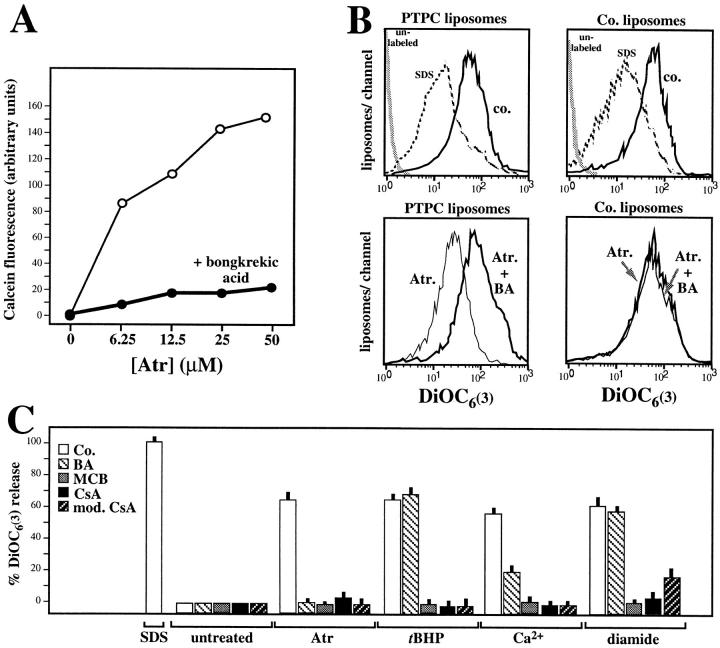
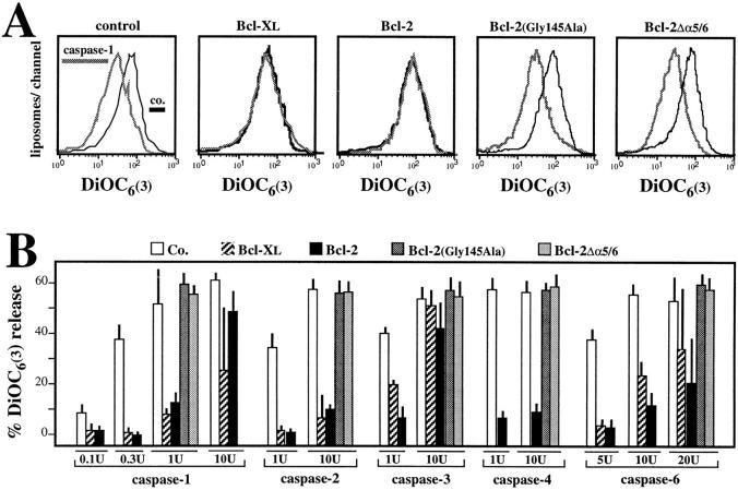
Early in programmed cell death (apoptosis), mitochondrial membrane permeability increases. This is at least in part due to opening of the permeability transition (PT) pore, a multiprotein complex built up at the contact site between the inner and the outer mitochondrial membranes. The PT pore has been previously implicated in clinically relevant massive cell death induced by toxins, anoxia, reactive oxygen species, and calcium overload. Here we show that PT pore complexes reconstituted in liposomes exhibit a functional behavior comparable with that of the natural PT pore present in intact mitochondria. The PT pore complex is regulated by thiol-reactive agents, calcium, cyclophilin D ligands (cyclosporin A and a nonimmunosuppressive cyclosporin A derivative), ligands of the adenine nucleotide translocator, apoptosis-related endoproteases (caspases), and Bcl-2-like proteins. Although calcium, prooxidants, and several recombinant caspases (caspases 1, 2, 3, 4, and 6) enhance the permeability of PT pore-containing liposomes, recombinant Bcl-2 or Bcl-XL augment the resistance of the reconstituted PT pore complex to pore opening. Mutated Bcl-2 proteins that have lost their cytoprotective potential also lose their PT modulatory capacity. In conclusion, the PT pore complex may constitute a crossroad of apoptosis regulation by caspases and members of the Bcl-2 family.


Figures

Figure 1
Figure 1
Enrichment of the PTPC. (A) Steps of the purification process. For details consult Materials and Methods. (B) Typical profile of an anion exchange chromatography performed on Triton-soluble proteins (A, 2). Hexokinase activity (solid line) elutes from the DE52 resin at a KCl concentration (linear gradient, dotted line) of 190 ± 10 mM. The most active fractions (bar) are recovered (A, 3) and reconstituted in liposomes as outlined in A. Cytochrome c elutes from the gradient with the major protein peak, at 70 ± 10 mM (arrow). (C) Typical profile of a molecular weight chromatography performed on liposomes reconstituted with the fractions recovered in B (A, 3). Note that hexokinase activity accumulates in a few fractions of the liposome–protein mixture. The fraction containing maximum hexokinase activity constitutes the PTPC liposomes (A, 4). (D) Immunochemical detection of proteins contained in PTPC. Proteins from successive steps of the purification procedure (A) were analyzed by Western blot for the presence of the indicated proteins. Proteins extracted from PTPC liposomes constitute the final step (A, 4) of the purification procedure. Results are representative for 22 (B and C), and two to three (D) independent determinations.
Figure 2
Figure 2
Two-dimensional gel electrophoresis of proteins extracted from PTPC liposomes (Fig. 1 A, 4). Silver-stained proteins whose abundance is consistently (three experiments) reduced upon digestion with caspase 1 (1.5 U/ml) are marked in rectangles. Results are representative for three independent experiments.
Figure 3
Figure 3
Function of reconstituted PTPC pores. (A) Release of calcein from PTPC-containing liposomes incubated with two antagonistic ANT ligands. Calcein-loaded PTPC liposomes were incubated with the indicated dose of Atr and/or bongkrekic acid (50 μM), followed by fluorometric determination of the calcein release into the supernatant. (B) Cytofluorometric profile of liposomes labeled with the potential-sensitive fluorochrome DiOC6(3). Liposomes were reconstituted either in the presence of the hexokinase-containing fraction (PTPC liposomes) or in its absence (control liposomes), treated with SDS (0.25%), Atr (25 μM), and/or bongkrekic acid (BA; 50 μM), followed by DiOC6(3) staining and cytofluorometric analysis. (C) PTPC liposomes treated with PT inducers (Atr [25 μM], CaCl2[25 μM], diamide [500 μM] or ter-butylhydroperoxide [tBHP, 500 μM]) and/or PT inhibitors (bongkrekic acid [BA; 50 μM], cyclosporin A [CsA; 10 μM], N-methyl-4-Val-CsA [mod. CsA; 10 μM], or monochlorobiman [MCB; 50 μM]). Results are expressed as percentage (X ± SD of triplicates) of the DiOC6(3) release induced by 0.25% SDS. Results are representative for at least three independent determinations.
Figure 4
Figure 4
Effects of Bcl-2 on PTPC. Hexokinase-enriched fractions (Fig. 1 A, 3) were incorporated into liposomes by dialysis in the presence or absence of recombinant Bcl-2, Bcl-2 (Gly145Ala), Bcl-2Δα5/6, or Bcl-XL, followed by functional analysis. (A) Representative fluorescence profiles of control PTPC and Bcl-2 PTPC liposomes treated with buffer only (control), SDS, or Atr, followed by incubation with DiOC6(3). Note the absence of Atr effects in Bcl-2 PTPC liposomes. (B) Incorporation of native and mutant Bcl-2 proteins into liposomes. Proteins were extracted from PTPC liposomes prepared in the presence or absence of the indicated Bcl-2 mutant, followed by immunochemical quantitation of Bcl-2 with a monoclonal antibody that recognizes an epitope (residues 20-34) not affected by the mutations. (C) Functional impact of Bcl-2 and Bcl-XL. The different PTPC liposome preparations were treated with Atr (25 μM), CaCl2 (25 μM), diamide (500 μM), or ter-butylhydroperoxide (tBHP, 500 μM) to determine the DiOC6(3) release. Results are representative for three to five independent experiments. 100% DiOC6(3) release was defined as the SDS-induced reduction of DiOC6(3) fluorescence observed in PTPC liposomes generated in the absence of Bcl-2 or Bcl-XL.
Figure 5
Figure 5
Effect of caspases on PTPC liposomes and isolated mitochondria. (A) Representative DiOC6(3) fluorescence histograms obtained after treatment of liposomes with various caspases (1.2 U/ml for caspase 1, 10 U/ml for caspase 6) in the presence or absence of the indicated caspase inhibitor (100 μM). (B) Dose dependency of effects obtained with different recombinant caspases on PTPC liposomes. (C) Effect of caspases on the Δψm. Mitochondria were treated during 30 min with 5 U caspase/200 μl, followed by determination of the Δψm using DiOC6(3). The protonophore m-chlorophenylhydrazone (50 μM) defined 100% Δψm disruption. (D) Release of AIF into the mitochondrial supernatant. Intact mitochondria were treated with the indicated caspase (5 U/200 μl), followed by centrifugation and removal of the supernatant that was tested for apoptogenic activity on isolated HeLa nuclei. The incubation was performed in the presence of tetrapeptide inhibitors (which inhibit caspases but not AIF) or in the presence of Z-VAD.fmk (which inhibits AIF) to exclude that nuclear DNA degradation is a direct caspase effect. Similar results were obtained with mouse and rat (not shown) hepatocyte mitochondria.
Figure 6
Figure 6
Effect of Bcl-2 on the caspase induced DiOC6(3) release observed in PTPC liposomes. (A) Representative DiOC6(3) staining profiles. Liposomes were generated in the presence of recombinant Bcl-2, Bcl-XL, and the indicated Bcl-2 mutants, treated with 1 U caspase 1, and labeled with DiOC6(3) to determine the DiOC6(3) release. Results are representative for at least three independent determinations. (B) Dose response curves of caspase effects on liposomes containing Bcl-XL, Bcl-2, or Bcl-2 mutants.
Figure 7
Figure 7
Cytochrome c retention in PTPC liposomes. Liposomes were generated in the absence or presence of recombinant Bcl-2, followed by generation of a KCl-dependent ion gradient and incorporation of cytochrome c during the sonication step. (A) Effect of SDS (0.25%), Atr (50 μM), or caspases 1 or 3 (1 U), as determined by flow cytometry after labeling with DiOC6(3). (B) Supernatants of the liposomes treated as in A were subjected to protein precipitation, followed by Western blot analysis of the release of cytochrome c. Note that the blot has been overexposed. The amount of cytochrome c released upon SDS treatment was estimated to be 1 μg, and the detection limit of the immunoblot is ∼10 ng/lane.

References

    1. Zamzami N, Marchetti P, Castedo M, Zanin C, Vayssière J-L, Petit PX, Kroemer G. Reduction in mitochondrial potential constitutes an early irreversible step of programmed lymphocyte death in vivo. J Exp Med. 1995;181:1661–1672. - PMC - PubMed
    1. Zamzami N, Marchetti P, Castedo M, Decaudin D, Macho A, Hirsch T, Susin SA, Petit PX, Mignotte B, Kroemer G. Sequential reduction of mitochondrial transmembrane potential and generation of reactive oxygen species in early programmed cell death. J Exp Med. 1995;182:367–377. - PMC - PubMed
    1. Kroemer G, Zamzami N, Susin SA. Mitochondrial control of apoptosis. Immunol Today. 1997;18:44–51. - PubMed
    1. Kroemer G. The proto-oncogene Bcl-2 and its role in regulating apoptosis. Nat Med. 1997;3:614–620. - PubMed
    1. Liu X, Kim CN, Yang J, Jemmerson R, Wang X. Induction of apoptotic program in cell-free extracts: requirement for dATP and cytochrome c. . Cell. 1996;86:147–157. - PubMed

Publication types