. 1998 Apr 20;187(8):1335-42.
doi: 10.1084/jem.187.8.1335.

Tolerization of anti-Galalpha1-3Gal natural antibody-forming B cells by induction of mixed chimerism

Affiliations

Tolerization of anti-Galalpha1-3Gal natural antibody-forming B cells by induction of mixed chimerism

Y G Yang et al. J Exp Med. .

Abstract

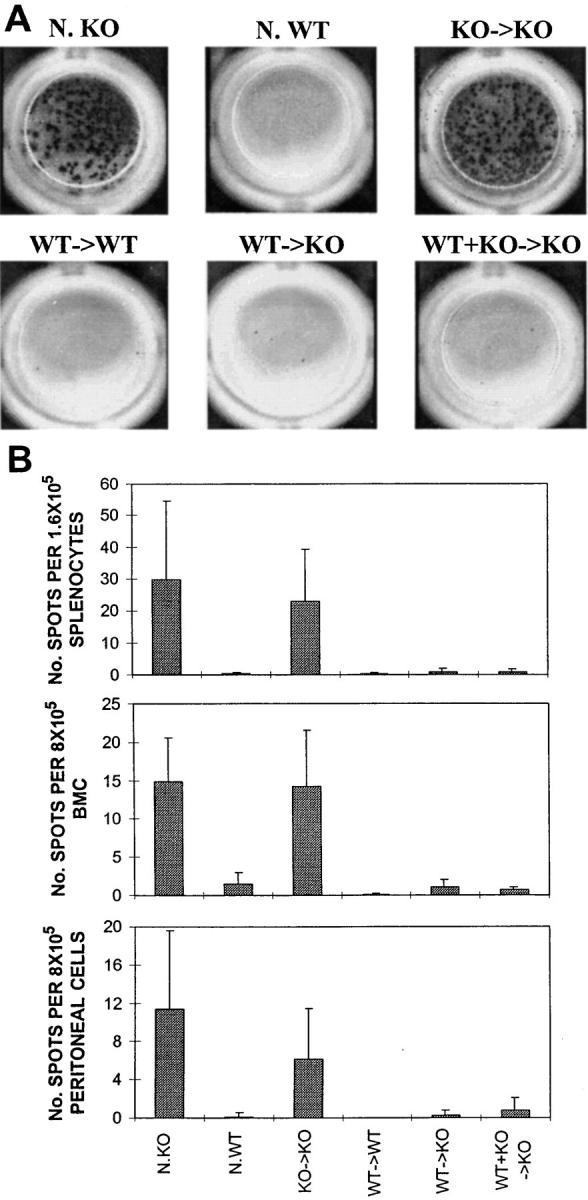
Xenotransplantation could overcome the severe shortage of allogeneic organs, a major factor limiting organ transplantation. Unfortunately, transplantation of organs from pigs, the most suitable potential donor species, results in hyperacute rejection in primate recipients, due to the presence of anti-Galalpha1-3Gal (Gal) natural antibodies (NAbs) in their sera. We evaluated the ability to tolerize anti-Gal NAb-producing B cells in alpha1,3-galactosyltransferase knockout (GalT KO) mice using bone marrow transplantation (BMT) from GalT+/+ wild-type (WT) mice. Lasting mixed chimerism was achieved in KO mice by cotransplantation of GalT KO and WT marrow after lethal irradiation. The levels of anti-Gal NAb in sera of mixed chimeras were reduced markedly 2 wk after BMT, and became undetectable at later time points. Immunization with Gal+/+ xenogeneic cells failed to stimulate anti-Gal antibody production in mixed chimeras, whereas the production of non-Gal-specific antixenoantigen antibodies was stimulated. An absence of anti-Gal-producing B cells was demonstrated by enzyme-linked immunospot assays in mixed KO + WT --> KO chimeras. Thus, mixed chimerism efficiently induces anti-Gal-specific B cell tolerance in addition to T cell tolerance, providing a single approach to overcoming both the humoral and the cellular immune barriers to discordant xenotransplantation.


Figures

Figure 1
Figure 1
Stable mixed chimerism in GalT KO recipients of mixed WT and KO BMC. PBLs were stained with anti–WT H-2Kb mAb 5F1-FITC and anti–CD19-PE or anti–CD4-PE plus anti–CD8-PE. Both WT (H-2bxd) and KO (H-2d) cells were detected in GalT KO recipients of mixed WT and KO BMC. (A) Representative FACS® profiles of PBLs from mixed and fully allogeneic chimeras at 12 wk after BMT. The two T cell populations of lower and higher intensity represent CD4+ and CD8+ T cells, respectively. (B) Percentages of WT B and T cells in the PBLs of untreated GalT KO mice (•, n = 3), WT mice (♦, n = 2), GalT KO mouse recipients of KO BMC (○, n = 3), WT BMC (□, n = 5), or mixed KO plus WT BMC (▵, n = 5), and WT mouse recipients of WT BMC (⋄, n = 3). Average (± SD) net percentages (after subtraction of background staining) of WT B cells (percentage 5F1+CD19+ cells/total B cells) and T cells (percentage 5F1+CD4/8+ cells/total T cells) are shown at the indicated time points. (C) BMC were prepared from GalT KO recipients of KO BMC (KOKO), WT BMC (WTKO), or WT plus KO BMC (WT+KOKO), or WT recipients of WT BMC (WTWT) 19 wk after BMT, and were stained by FITC-conjugated anti–WT H-2Kb mAb 5F1, anti–CD19-PE, and anti–CD4-Bio plus anti–CD8-Bio, followed by incubation with Cy-chrome-streptavidin. Typical histogram representation of WT (5F1+) cells on gated B cells (CD19+, top), T cells (CD4+/CD8+, middle), and nonlymphoid cells (CD19CD4CD8, bottom [most of these were Mac-1+]) are shown. Similar results were observed for spleen and intraperitoneal cells (data not shown).
Figure 2
Figure 2
Serum levels of anti-Gal NAb in lethally irradiated GalT KO mice. Peripheral blood samples (40 μl) were collected from lethally irradiated (9.75 Gy) GalT KO mice that did (⋄) or did not (•) receive TCD GalT KO BMC (5 × 10⁶ cells/mouse), and from 9.75 Gy–irradiated WT mice (□) at the indicated times, and serum levels of anti-Gal NAb were measured by FCM. WT scid mouse cells were stained with 10 μl serum, and NAbs were detected using rat anti–mouse IgM-FITC as secondary mAb. The anti-Gal NAb (NAB) levels are presented as median fluorescence intensity (MFI). Each line represents an individual animal.
Figure 3
Figure 3
Reduced anti-Gal NAb levels in sera of mixed WT+KO→ KO chimeras. (A) Representative histograms obtained by FCM analysis show an absence of anti-Gal NAb in mixed and fully WT→ KO chimeras. WT RAG-1−/− mouse cells were stained with sera from normal KO and WT mice and BMT recipients, and NAbs were detected using rat anti–mouse IgM-FITC as secondary mAb. Typical histogram appearances for normal KO and WT mice, and for BMT recipients at 4 wk after BMT are shown. (B) Kinetics of serum anti-Gal NAb levels measured by Gal-specific ELISA assay. Sera were collected from normal KO and WT control mice and BMT recipients at 2, 4, 8, and 12 wk after BMT, and anti-Gal NAb (IgM plus IgG) levels were determined. Average and SDs for the individual groups are shown. Number of animals in each group: normal KO (x), 3; normal WT (▪), 2; KO→ KO (•), 3; WT→ WT (□), 3; WT→ KO (▴), 5; WT+KO→ KO (♦), 5.
Figure 4
Figure 4
Reduced number of B cells producing anti-Gal antibodies (IgM plus IgG) in mixed chimeras. Spleen cells, BMC, and intraperitoneal cells prepared from BMT recipients or normal (N.) KO or WT mice 8 d after sensitization with rabbit RBC were serially diluted and used in ELISPOT assays. (A) Typical ELISPOT wells of spleen cells from indicated mice. (B) Average number of spots (± SD) (anti-Gal Ig–producing B cells) in spleen (top), BMC (middle), and intraperitoneal cells (bottom) of the mice killed 8 d after rabbit RBC immunization.
Figure 5
Figure 5
Absence of anti-Gal antibodies in sera of xenogeneic cell–sensitized mixed chimeras, and evidence of sensitization to xenoantigens. (A) Serum levels of anti–rabbit and anti-Gal antibodies in rabbit RBC–sensitized mice. Anti–rabbit RBCs and anti-Gal antibodies were detected by staining rabbit RBC and WT scid mouse cells, respectively, with serially diluted serum, followed by incubation with FITC-conjugated anti–mouse IgM. Left column, Rabbit RBCs stained with sera of indicated mice collected before (Pre) (10 μl of undiluted serum) and 8 d after (Post) sensitization (10 μl of twofold diluted serum). Right column, WT scid cells stained with sera collected 8 d after sensitization with rabbit RBC (10 μl of 50-fold diluted serum). Sera from scid mice (N. SCID) were used as negative controls. (B and C) Serum levels of anti–pig and anti-Gal antibodies in pig PBMC–sensitized mice. Lethally irradiated BMT recipients of KO→ KO, WT→ KO, or WT+KO→ KO and normal GalT KO mice were immunized three times by intraperitoneal injection of 106 pig PBMCs at 15, 16, and 22 wk after BMT, and sera were collected 3 wk after the last injection. Sera from unimmunized normal WT (N. WT) and GalT KO (N. KO) mice were used as controls. (B) Serum levels of anti–pig PBMC IgM measured by FCM analysis. Pig PBMCs were stained with sera (10 μl of undiluted serum) collected from indicated groups (four mice for each group), and anti–pig IgM was detected using FITC-conjugated anti– mouse IgM. The anti–pig PBMC IgM levels are presented as median fluorescence intensity (MFI). (C) Serum levels of anti-Gal antibodies in pig PBMC-sensitized normal GalT KO mice (n = 4; ♦, dotted line) and BMT recipients of KO→ KO (n = 5; •, solid line), WT→ KO (n = 6; ▪, dotted line), or WT+KO→ KO (n = 8; ▴, solid line) detected by ELISA assay. Average and SDs for the individual groups are shown.

References

    1. Cooper DKC. Xenografting: how great is the clinical need? . Xeno. 1993;2:25–26.
    1. Platt JL. Xenotransplantation: recent progress and current perspectives. Curr Opin Immunol. 1996;8:721–728. - PubMed
    1. Sachs DH. The pig as a xenograft donor. Pathol Biol. 1994;42:217–219. - PubMed
    1. Cooper, D.K.C., Y. Ye, L.L. Rolf, Jr., and N. Zuhdi. 1991. The pig as potential organ donor for man. In Xenotransplantation. D.K.C. Cooper, E. Kemp, K. Reemtsma, and D.J.G. White, editors. Springer-Verlag, Heidelberg. 481–500.
    1. Auchincloss HJ. Xenogeneic transplantation. A review. Transplantation (Baltimore) 1988;46:1–20. - PubMed

Publication types

Substances