. 1998 Apr 20;187(8):1343-8.
doi: 10.1084/jem.187.8.1343.

A double-edged kinase Lyn: a positive and negative regulator for antigen receptor-mediated signals

Affiliations

A double-edged kinase Lyn: a positive and negative regulator for antigen receptor-mediated signals

H Nishizumi et al. J Exp Med. .

Abstract

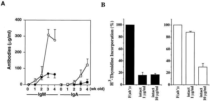
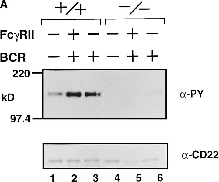
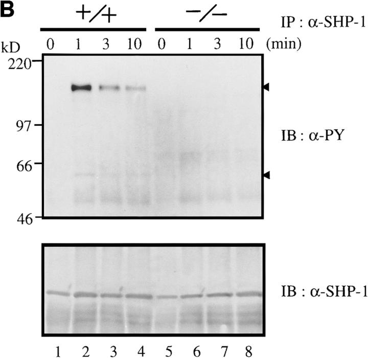
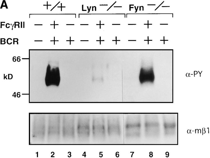
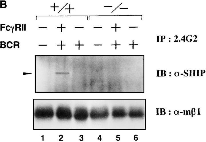
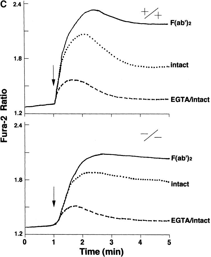
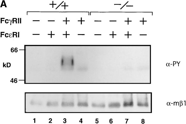
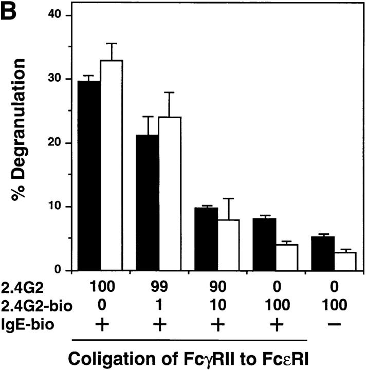
B cells from young lyn-/- mice are hyperresponsive to anti-IgM-induced proliferation, suggesting involvement of Lyn in negative regulation of B cell antigen receptor (BCR)-mediated signaling. Here we show that tyrosine phosphorylation of FcgammaRIIB and CD22 coreceptors, which are important for feedback suppression of BCR-induced signaling, was severely impaired in lyn-/- B cells upon their coligation with the BCR. Hypophosphorylation on tyrosine residues of these molecules resulted in failure of recruiting the tyrosine phosphatase SHP-1 and inositol phosphatase SHIP, SH2-containing potent inhibitors of BCR-induced B cell activation, to the coreceptors. Consequently, lyn-/- B cells exhibited defects in suppressing BCR-induced Ca2+ influx and proliferation. Thus, Lyn is critically important in tyrosine phosphorylation of the coreceptors, which is required for feedback suppression of B cell activation.


Figures

Figure 1
Figure 1
(A) Serum levels of IgM and IgA in unimmunized infant mice. Wild-type mice (closed circles) and lyn −/− mice (open circles) were bled at 0–4 wk of age. Concentrations of IgM and IgA in their serum were determined by isotype-specific ELISA (13). Averages of the results from three experiments are shown. (B) Relative proliferative responses to BCR cross-linking. Splenic B cells from wild-type (left) or lyn −/− (right) mice were cultured with goat F(ab′)2 anti-IgM (5 μg/ml) or goat intact anti-IgM (1 or 10 μg/ml). After 42 h of incubation, cells were pulse labeled for 6 h with [3H]thymidine, and [3H]thymidine incorporation was determined (13). All assays were performed in triplicate. The proliferative responses were presented with [3H]thymidine incorporation expressed as a percentage of the response against F(ab′)2 anti-IgM stimulation.
Figure 2
Figure 2
(A) Anti-IgM–induced tyrosine phosphorylation of CD22. Splenic B cells were stimulated with F(ab′)2 anti-IgM (30 μg/ml) (lanes 3 and 6) or intact anti-IgM (50 μg/ml) (lanes 2 and 5). Anti-CD22 immunoprecipitates from the cell lysates were probed with α-PY (phosphotyrosine antibody; top) or anti-CD22 antibody (bottom) by immunoblotting. (B) Anti-IgM–induced tyrosine phosphorylation of SHP-1 and its associated molecules. Splenic B cells were stimulated with intact anti-IgM (50 μg/ml) for indicated times. Anti–SHP-1 immunoprecipitates from the cell lysates were probed with antiphosphotyrosine monoclonal antibody 4G10 (α-PY) by immunoblotting. About 150-kD proteins (upper arrowhead) coimmunoprecipitated with SHP-1 (lower arrowhead) corresponded to CD22.
Figure 2
Figure 2
(A) Anti-IgM–induced tyrosine phosphorylation of CD22. Splenic B cells were stimulated with F(ab′)2 anti-IgM (30 μg/ml) (lanes 3 and 6) or intact anti-IgM (50 μg/ml) (lanes 2 and 5). Anti-CD22 immunoprecipitates from the cell lysates were probed with α-PY (phosphotyrosine antibody; top) or anti-CD22 antibody (bottom) by immunoblotting. (B) Anti-IgM–induced tyrosine phosphorylation of SHP-1 and its associated molecules. Splenic B cells were stimulated with intact anti-IgM (50 μg/ml) for indicated times. Anti–SHP-1 immunoprecipitates from the cell lysates were probed with antiphosphotyrosine monoclonal antibody 4G10 (α-PY) by immunoblotting. About 150-kD proteins (upper arrowhead) coimmunoprecipitated with SHP-1 (lower arrowhead) corresponded to CD22.
Figure 3
Figure 3
(A) Anti-IgM–induced tyrosine phosphorylation of FcγRIIB. Splenic B cells from wild-type (lanes 1–3), lyn −/− (lanes 4–6), or fyn −/− (lanes 7–9) mice were stimulated with F(ab′)2 anti-IgM (30 μg/ml; lanes 3, 6, and 9) or intact anti-IgM (50 μg/ml; lanes 2, 5, and 8) for 2 min. Proteins were immunoprecipitated with 2.4G2 anti-FcγRIIB monoclonal antibody from the cell lysates, and the precipitates probed with α-PY (top) or anti-FcγRIIB antibody (α-mβ1; bottom) by immunoblotting. (B) Recruitment of SHIP to FcγRIIB. Splenic B cells from wild-type (lanes 1–3) or lyn −/− (lanes 4–6) mice were stimulated with F(ab′)2 anti-IgM (30 μg/ml; lanes 3 and 6) or intact anti-IgM (50 μg/ml; lanes 2 and 5) for 2 min. Anti-FcγRIIB (2.4G2) immunoprecipitates from the cell lysates were probed with anti-SHIP antibodies by immunoblotting. (C) Time course of Ca2+ flux in splenic B cells upon BCR coligation to FcγRIIB. Panels show real time Fura-2 ratios (340/380) for splenic B cells of wild-type mice (top) and lyn −/− mice (bottom). Splenic B cells were stimulated with F(ab′)2 anti-IgM (5 μg/ml; line), intact anti-IgM (10 μg/ ml; dotted line), or intact anti-IgM (10 μg/ml) in the presence of 1.5 mM EGTA (perforated line).
Figure 3
Figure 3
(A) Anti-IgM–induced tyrosine phosphorylation of FcγRIIB. Splenic B cells from wild-type (lanes 1–3), lyn −/− (lanes 4–6), or fyn −/− (lanes 7–9) mice were stimulated with F(ab′)2 anti-IgM (30 μg/ml; lanes 3, 6, and 9) or intact anti-IgM (50 μg/ml; lanes 2, 5, and 8) for 2 min. Proteins were immunoprecipitated with 2.4G2 anti-FcγRIIB monoclonal antibody from the cell lysates, and the precipitates probed with α-PY (top) or anti-FcγRIIB antibody (α-mβ1; bottom) by immunoblotting. (B) Recruitment of SHIP to FcγRIIB. Splenic B cells from wild-type (lanes 1–3) or lyn −/− (lanes 4–6) mice were stimulated with F(ab′)2 anti-IgM (30 μg/ml; lanes 3 and 6) or intact anti-IgM (50 μg/ml; lanes 2 and 5) for 2 min. Anti-FcγRIIB (2.4G2) immunoprecipitates from the cell lysates were probed with anti-SHIP antibodies by immunoblotting. (C) Time course of Ca2+ flux in splenic B cells upon BCR coligation to FcγRIIB. Panels show real time Fura-2 ratios (340/380) for splenic B cells of wild-type mice (top) and lyn −/− mice (bottom). Splenic B cells were stimulated with F(ab′)2 anti-IgM (5 μg/ml; line), intact anti-IgM (10 μg/ ml; dotted line), or intact anti-IgM (10 μg/ml) in the presence of 1.5 mM EGTA (perforated line).
Figure 4
Figure 4
(A) Tyrosine phosphorylation of FcγRIIB upon FcεRI and FcγRIIB coligation. BMMCs from wild-type (lanes 1–4), lyn −/− (lanes 5–8) mice were sensitized with anti-DNP monoclonal IgE (10 μg/ml) followed by stimulation for 2 min at 37°C with 30 ng/ml DNP-HSA (FcεRI cross-linking; lanes 2 and 6), or DNP-HSA/rabbit anti-HSA IgG immune complexes (FcγRII coligation to FcεRI; lanes 3 and 7). BMMCs were stimulated with DNP-HSA/rabbit anti-HSA IgG immune complexes without IgE sensitization (lanes 4 and 8). Anti-FcγRIIB (2.4G2) immunoprecipitates from the cell lysates were probed with α-PY (top) or anti-FcγRIIB antibody (α-mβ1) (bottom) by immunoblotting. (B) Inhibitory effect of FcγRIIB coligation to FcεRI in lyn −/− BMMCs. The mast cell was sensitized with biotinylated mouse IgE, followed by cross-linking with streptavidin (no FcγRII coligation). FcγRIIB was coligated to FcεRI by adding biotinylated anti-FcγRII monoclonal antibody (biotin-2.4G2) at the sensitization step (100% FcγRII coligation). A mixture of biotinylated/nonbiotinylated 2.4G2 (1/9 for 10% or 1/99 for 1% coligation) was used to vary the extent of FcγRII coligation. The degree of degranulation was determined by measuring the release of β-hexosaminidase as described (12). Closed columns, the results of wild-type mice; open columns, the results of lyn −/− mice. Standard errors of triplicate samples are indicated on each column.
Figure 4
Figure 4
(A) Tyrosine phosphorylation of FcγRIIB upon FcεRI and FcγRIIB coligation. BMMCs from wild-type (lanes 1–4), lyn −/− (lanes 5–8) mice were sensitized with anti-DNP monoclonal IgE (10 μg/ml) followed by stimulation for 2 min at 37°C with 30 ng/ml DNP-HSA (FcεRI cross-linking; lanes 2 and 6), or DNP-HSA/rabbit anti-HSA IgG immune complexes (FcγRII coligation to FcεRI; lanes 3 and 7). BMMCs were stimulated with DNP-HSA/rabbit anti-HSA IgG immune complexes without IgE sensitization (lanes 4 and 8). Anti-FcγRIIB (2.4G2) immunoprecipitates from the cell lysates were probed with α-PY (top) or anti-FcγRIIB antibody (α-mβ1) (bottom) by immunoblotting. (B) Inhibitory effect of FcγRIIB coligation to FcεRI in lyn −/− BMMCs. The mast cell was sensitized with biotinylated mouse IgE, followed by cross-linking with streptavidin (no FcγRII coligation). FcγRIIB was coligated to FcεRI by adding biotinylated anti-FcγRII monoclonal antibody (biotin-2.4G2) at the sensitization step (100% FcγRII coligation). A mixture of biotinylated/nonbiotinylated 2.4G2 (1/9 for 10% or 1/99 for 1% coligation) was used to vary the extent of FcγRII coligation. The degree of degranulation was determined by measuring the release of β-hexosaminidase as described (12). Closed columns, the results of wild-type mice; open columns, the results of lyn −/− mice. Standard errors of triplicate samples are indicated on each column.

Similar articles

Cited by

References

    1. Burkhardt AL, Brunswick M, Bolen JB, Mond JJ. Anti-immunoglobulin stimulation of B lymphocytes activates src-related protein kinases. Proc Natl Acad Sci USA. 1991;88:7410–7414. - PMC - PubMed
    1. Yamanashi Y, Kakiuchi T, Mizuguchi J, Yamamoto T, Toyoshima K. Association of B cell antigen receptor with protein tyrosine kinase Lyn. Science. 1991;251:192–194. - PubMed
    1. Eisenman E, Bolen JB. Engagement of the high-affinity IgE receptor activates src-protein–related tyrosine kinases. Nature. 1992;355:78–80. - PubMed
    1. Kurosaki T, Takata M, Yamanashi Y, Inazu T, Taniguchi T, Yamamoto T, Yamamura H. Syk activation by the Src-family tyrosine kinase in the B cell receptor signaling. J Exp Med. 1994;179:1725–1729. - PMC - PubMed
    1. Sidorenko SP, Law C-L, Chandran KA, Clark EA. Human spleen tyrosine kinase p72Syk associates with the Src-family p53/56Lynand a 120-kDa phosphoprotein. Proc Natl Acad Sci USA. 1995;92:359–363. - PMC - PubMed

Publication types