. 1998 Apr 20;141(2):309-19.
doi: 10.1083/jcb.141.2.309.

Centromere protein B null mice are mitotically and meiotically normal but have lower body and testis weights

Affiliations

Centromere protein B null mice are mitotically and meiotically normal but have lower body and testis weights

D F Hudson et al. J Cell Biol. .

Abstract

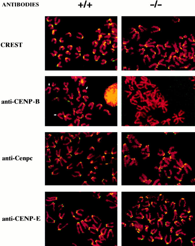
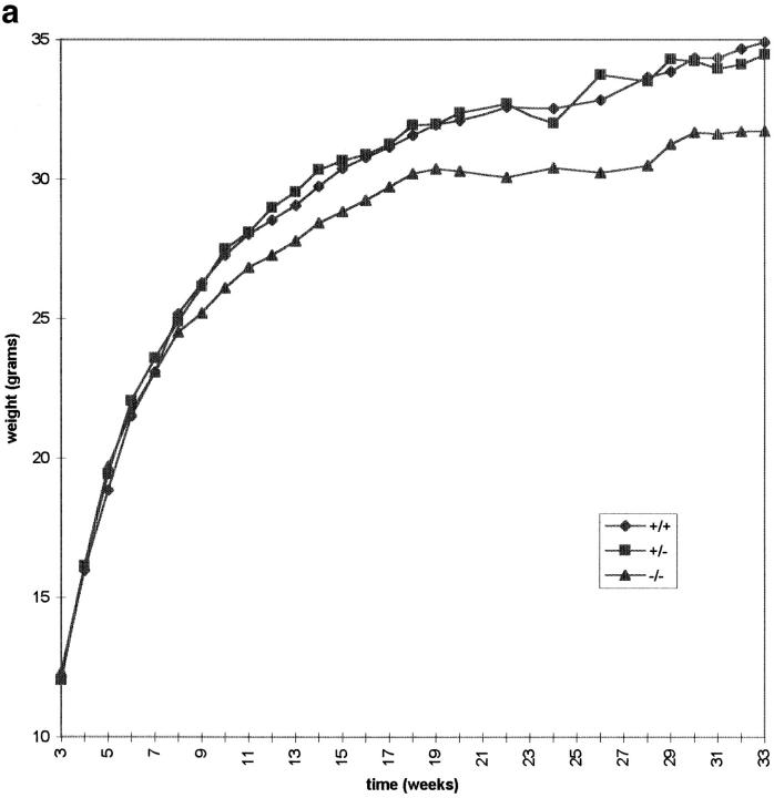
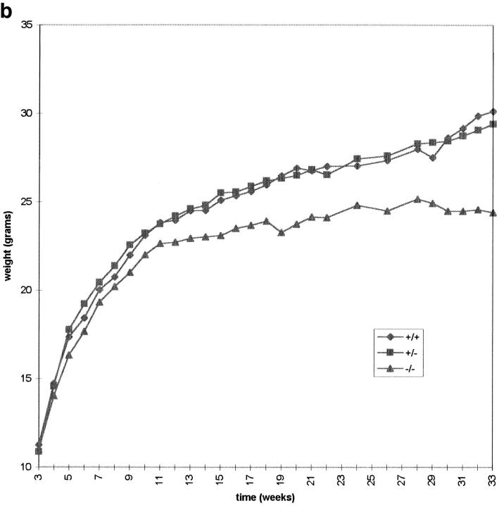
CENP-B is a constitutive centromere DNA-binding protein that is conserved in a number of mammalian species and in yeast. Despite this conservation, earlier cytological and indirect experimental studies have provided conflicting evidence concerning the role of this protein in mitosis. The requirement of this protein in meiosis has also not previously been described. To resolve these uncertainties, we used targeted disruption of the Cenpb gene in mouse to study the functional significance of this protein in mitosis and meiosis. Male and female Cenpb null mice have normal body weights at birth and at weaning, but these subsequently lag behind those of the heterozygous and wild-type animals. The weight and sperm content of the testes of Cenpb null mice are also significantly decreased. Otherwise, the animals appear developmentally and reproductively normal. Cytogenetic fluorescence-activated cell sorting and histological analyses of somatic and germline tissues revealed no abnormality. These results indicate that Cenpb is not essential for mitosis or meiosis, although the observed weight reduction raises the possibility that Cenpb deficiency may subtly affect some aspects of centromere assembly and function, and result in reduced rate of cell cycle progression, efficiency of microtubule capture, and/or chromosome movement. A model for a functional redundancy of this protein is presented.


Figures

Figure 1
Figure 1
Cenpb targeting construct and screening strategies. (a) Intronless wild-type Cenpb locus showing the start and stop codons of the 1.8-kb coding region (solid box). The positions of the NH2-terminal 125–amino acid centromere DNA-binding domain and the COOH-terminal 59– amino acid dimerization domain are indicated (stippled boxes). Numbers below the line indicate nucleotide positions of the Cenpb sequence (EMBL accession no. X55038). (b) The IRES(neo) and IRES(hygro) targeting constructs, showing a 5480-bp contiguous KspI/HindIII Cenpb fragment that is interrupted by a frame shift-linker (D/TAA) containing stop codons and a new DraI site within the centromere DNA-binding domain, and an IRES(neo or hygro) cassette within the 3′ untranslated region. The H-A-S region on the right is vector DNA. Solid crosses denote the preferred regions of homologous recombination that result in the desired gene disruption event shown in c, whereas the broken cross indicates an undesired alternative region of recombination which, together with that at the solid-cross region to the right, results in incorporation of the IRES(neo or hygro) cassette, but not the D/TAA linker, generating a targeted event that is not accompanied by a gene disruption. p(A), polyadenylation signal; D, DraI; K, KspI; H, HindIII; A, AatII; S, SspI. (c) Correctly targeted Cenpb allele. (d) Screening strategies for targeted events. In Southern analysis of DraI digests, the 5′-probe (open box) detects a wild-type allele (+) of 5,300 bp, a desired targeted gene disruption allele (−) of 700 bp, and an undesired targeted but undisrupted allele (o) of 7,000 bp. Fd-1 and Rev-2/Rev-3 are PCR primers flanking the D/TAA linker region used for mouse tail DNA genotyping and RT-PCR. (e) Southern blot screening of transfected ES cells digested with DraI using the 5′-probe. Lane 5 shows a heterozygous colony with the desired gene disruption event. (f) Southern blot screening of tail DNA from mouse progeny of a +/− × +/− cross, digested with DraI and probed with the 5′-probe, showing the detection of homozygous Cenpb disruption (lane 1), heterozygous mice (lanes 2 and 3), and wild-type animals (lanes 4 and 5). (g) RT-PCR of total RNA from +/+ R1, +/− IRES(neo)-targeted R1-26, and −/− IRES(neo) and IRES(hygro) double-targeted R1-189N/H cells using the Fd-1/Rev-2 (lanes 1–3) and Fd-1/Rev-3 (lanes 4–7) primer sets. Incorporation of the D/TAA linker in the targeted allele increases the PCR products for both primer sets by 26 bp compared with the wild-type allele. Only the larger band containing the D/TAA linker was detected in the −/− cells (lanes 3 and 6). Lane 7, PCR control with no RNA.
Figure 1
Figure 1
Cenpb targeting construct and screening strategies. (a) Intronless wild-type Cenpb locus showing the start and stop codons of the 1.8-kb coding region (solid box). The positions of the NH2-terminal 125–amino acid centromere DNA-binding domain and the COOH-terminal 59– amino acid dimerization domain are indicated (stippled boxes). Numbers below the line indicate nucleotide positions of the Cenpb sequence (EMBL accession no. X55038). (b) The IRES(neo) and IRES(hygro) targeting constructs, showing a 5480-bp contiguous KspI/HindIII Cenpb fragment that is interrupted by a frame shift-linker (D/TAA) containing stop codons and a new DraI site within the centromere DNA-binding domain, and an IRES(neo or hygro) cassette within the 3′ untranslated region. The H-A-S region on the right is vector DNA. Solid crosses denote the preferred regions of homologous recombination that result in the desired gene disruption event shown in c, whereas the broken cross indicates an undesired alternative region of recombination which, together with that at the solid-cross region to the right, results in incorporation of the IRES(neo or hygro) cassette, but not the D/TAA linker, generating a targeted event that is not accompanied by a gene disruption. p(A), polyadenylation signal; D, DraI; K, KspI; H, HindIII; A, AatII; S, SspI. (c) Correctly targeted Cenpb allele. (d) Screening strategies for targeted events. In Southern analysis of DraI digests, the 5′-probe (open box) detects a wild-type allele (+) of 5,300 bp, a desired targeted gene disruption allele (−) of 700 bp, and an undesired targeted but undisrupted allele (o) of 7,000 bp. Fd-1 and Rev-2/Rev-3 are PCR primers flanking the D/TAA linker region used for mouse tail DNA genotyping and RT-PCR. (e) Southern blot screening of transfected ES cells digested with DraI using the 5′-probe. Lane 5 shows a heterozygous colony with the desired gene disruption event. (f) Southern blot screening of tail DNA from mouse progeny of a +/− × +/− cross, digested with DraI and probed with the 5′-probe, showing the detection of homozygous Cenpb disruption (lane 1), heterozygous mice (lanes 2 and 3), and wild-type animals (lanes 4 and 5). (g) RT-PCR of total RNA from +/+ R1, +/− IRES(neo)-targeted R1-26, and −/− IRES(neo) and IRES(hygro) double-targeted R1-189N/H cells using the Fd-1/Rev-2 (lanes 1–3) and Fd-1/Rev-3 (lanes 4–7) primer sets. Incorporation of the D/TAA linker in the targeted allele increases the PCR products for both primer sets by 26 bp compared with the wild-type allele. Only the larger band containing the D/TAA linker was detected in the −/− cells (lanes 3 and 6). Lane 7, PCR control with no RNA.
Figure 2
Figure 2
Immunostaining of centromere proteins (yellow signals) in +/+ R1 and −/− R1-189N/H cell lines using anticentromere antibodies. Results for the +/− R1-26 cell line were similar to those for the +/+ cell line and are not shown. Uniform signals were observed in all the centromeres in both cell lines when stained with CREST, anti-Cenpc, and anti-CENP-E antibodies. Note differences in the intensity of anti-CENP-B staining on different chromosomes in the +/+ cell line, with some centromeres (arrows) showing little or no detectable signals. No Cenpb signal was seen on the centromeres of the −/− cell line, even after maximal enhancement of fluorescence signal (thus the paler background) using computer imaging facility.
Figure 3
Figure 3
Total body weight of male (a) and female (b) IRES(neo) mice. The male data were collected from an average of 33, 46, and 19 animals, whereas the female data were from an average of 24, 39, and 17 animals for each time point for the +/+, +/−, and −/− genotypes, respectively. Individual weight value, measured weekly or fortnightly, represents the means of the weights for the total number of animals used at that time point.
Figure 3
Figure 3
Total body weight of male (a) and female (b) IRES(neo) mice. The male data were collected from an average of 33, 46, and 19 animals, whereas the female data were from an average of 24, 39, and 17 animals for each time point for the +/+, +/−, and −/− genotypes, respectively. Individual weight value, measured weekly or fortnightly, represents the means of the weights for the total number of animals used at that time point.
Figure 4
Figure 4
Model depicting the role of CENP-B and its putative functional homolog CENP-Z. (a) A centromeric satellite DNA array, showing individual monomers containing either a CENP-B-box (red bar) or a CENP-Z-box motif (blue bar). (b) CENP-B proteins (red circles) bind to CENP-B-box motifs and undergo dimerization to cross-link the array into a more stable higher order configuration. This mode of organization is presumably found on chromosomes where CENP-B boxes are prevalent, including the human and mouse autosomes and X chromosomes. (c) In the absence of CENP-B-binding, a functionally related protein CENP-Z (blue circle) assumes the role of CENP-B to cross-link CENP-Z box–containing monomers. This mode of organization is suggested for the human and mouse Y chromosomes, various CENP-B box-poor centromeric subdomains, and the centromeres of African green monkey and Cenpb null mice.

References

    1. Alexandrov IA, Medvedev LI, Mashkova TD, Kisselev LL, Romanova LY, Yurov YB. Definition of a new alpha satellite suprachromosomal family characterized by monomeric organization. Nucl Acids Res. 1993;21:2209–2215. - PMC - PubMed
    1. Bejarano LA, Valdivia MM. Molecular cloning of an intronless gene for the hamster centromere antigen CENP-B. Biochim Biophys Acta. 1996;1307:21–25. - PubMed
    1. Bernat RL, Borisy GG, Rothfield NF, Earnshaw WC. Injection of anticentromere antibodies in interphase disrupts events required for chromosome movement in mitosis. J Cell Biol. 1990;111:1519–1533. - PMC - PubMed
    1. Bernat RL, Delannoy MR, Rothfield NF, Earnshaw WC. Disruption of centromere assembly during interphase inhibits kinetochore morphogenesis and function in mitosis. Cell. 1991;66:1229–1238. - PubMed
    1. Brenner S, Pepper D, Berns MW, Tan E, Brinkley BR. Kinetochore structure, duplication and distribution in mammalian cells: analysis by human autoantibodies from scleroderma patients. J Cell Biol. 1981;91:95–102. - PMC - PubMed

Publication types