. 1998 Apr 20;141(2):321-33.
doi: 10.1083/jcb.141.2.321.

Human autoantibodies reveal titin as a chromosomal protein

Affiliations

Human autoantibodies reveal titin as a chromosomal protein

C Machado et al. J Cell Biol. .

Abstract

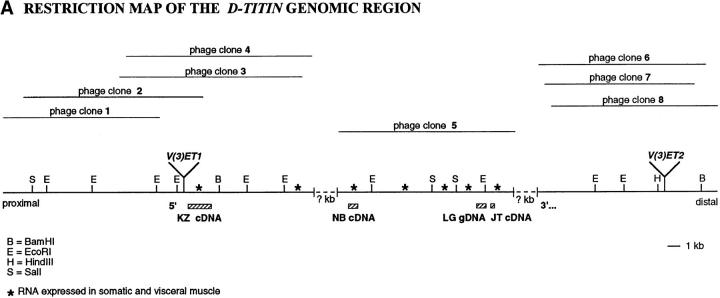
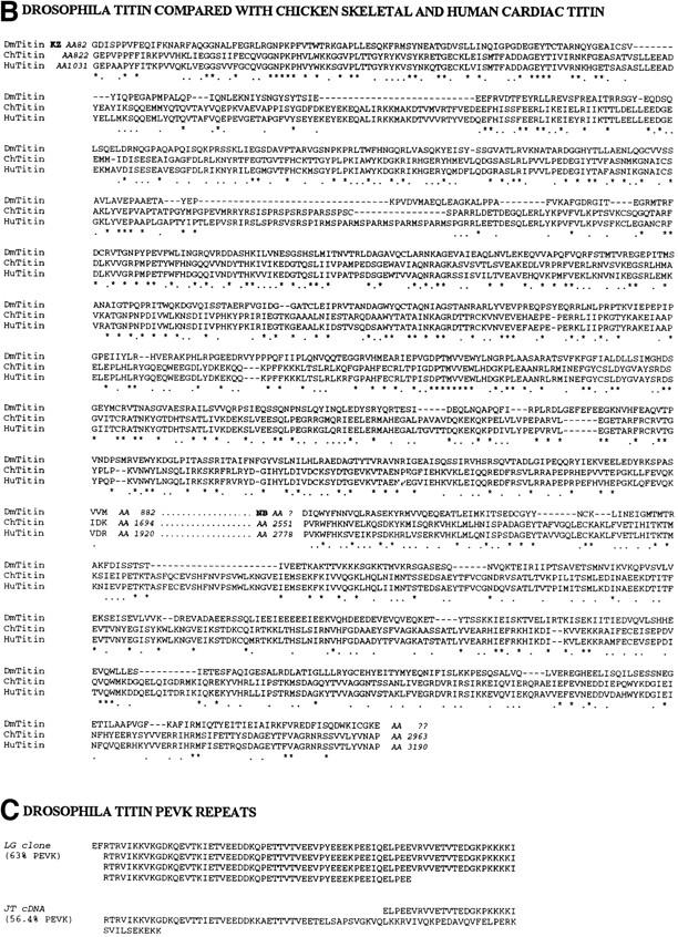
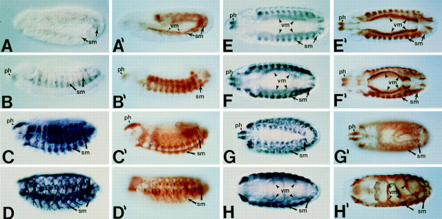
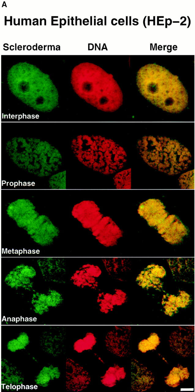
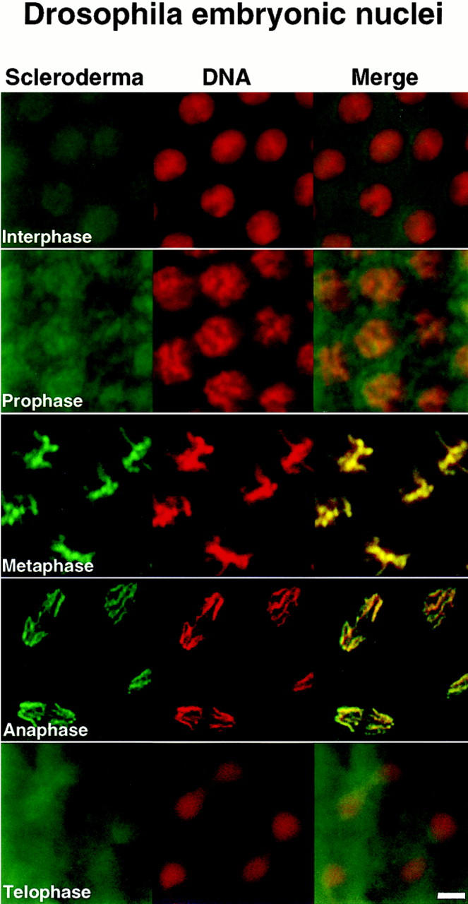
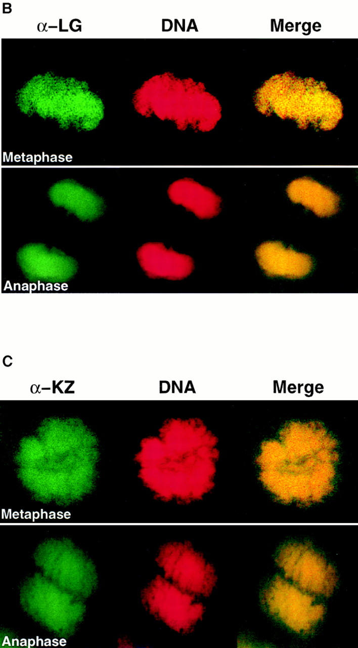
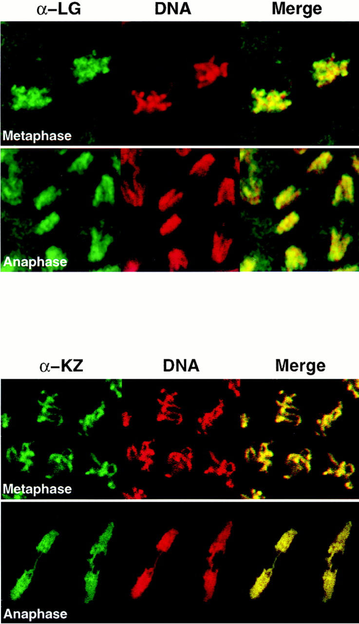
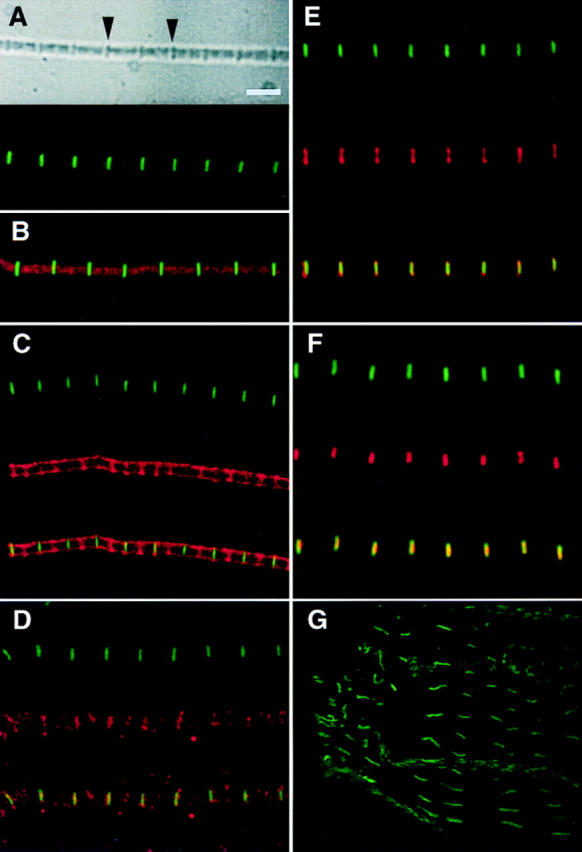
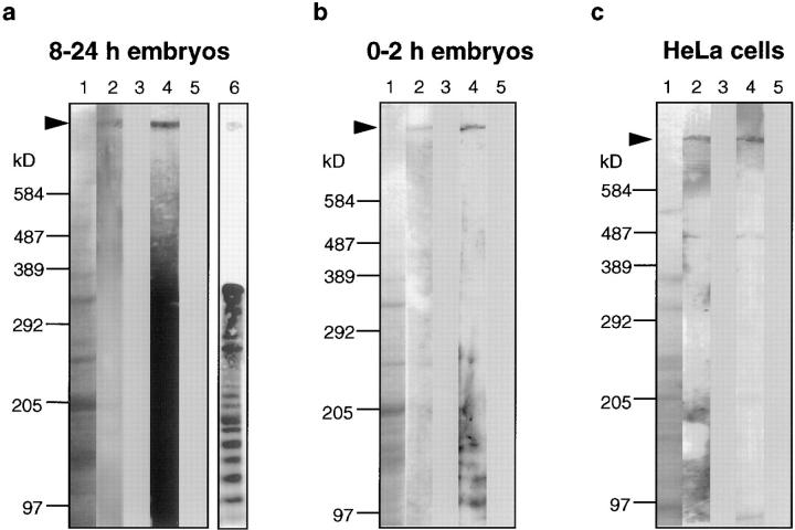
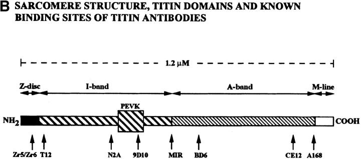
Assembly of the higher-order structure of mitotic chromosomes is a prerequisite for proper chromosome condensation, segregation and integrity. Understanding the details of this process has been limited because very few proteins involved in the assembly of chromosome structure have been discovered. Using a human autoimmune scleroderma serum that identifies a chromosomal protein in human cells and Drosophila embryos, we cloned the corresponding Drosophila gene that encodes the homologue of vertebrate titin based on protein size, sequence similarity, developmental expression and subcellular localization. Titin is a giant sarcomeric protein responsible for the elasticity of striated muscle that may also function as a molecular scaffold for myofibrillar assembly. Molecular analysis and immunostaining with antibodies to multiple titin epitopes indicates that the chromosomal and muscle forms of titin may vary in their NH2 termini. The identification of titin as a chromosomal component provides a molecular basis for chromosome structure and elasticity.


Figures

Figure 2
Figure 2
The chromosome-associated protein is Drosophila TITIN. (A) Restriction map of the genomic region of the D-Titin gene. The genomic phage clones were either isolated directly (phage clone 5) using the LG genomic DNA expression clone as a probe, or were isolated using DNA flanking two nearby P-element insertions. Phage clones 1–4 and 6–8 were isolated with DNA flanking the v(3)ET1 and v(3)ET2 insertions, respectively. Three different D-Titin cDNA fragments were isolated from multiple independent screens of seven available cDNA libraries. The 5′ KZ cDNA was isolated from a 9–12 h embryonic cDNA library (Zinn et al., 1988). We infer that the KZ cDNA encodes an NH2 terminus based on the presence of a putative initiator methionine codon followed by an ORF encoding 882 AA. The ORF is flanked at the 5′ end by 389 nt of noncoding sequence. The NB cDNA was isolated from a 12–24 h embryonic cDNA library (Brown and Kafatos, 1988). Within the unprocessed NB cDNA, there is a 1-kb ORF flanked at its 5′ end by a 3′ splice acceptor site and at its 3′ end by a 5′ splice donor site (Mount, 1982; Mount et al., 1992). Several small (⩽312 nt) cDNAs were isolated from a 0–24 h embryonic cDNA library (Tamkun et al., 1991). The largest cDNA isolated from the Tamkum library is indicated as JT cDNA. Multiple unsuccessful attempts were made to connect the genomic DNA from phage clone 5 to the surrounding phage containing DNA from this region. Nonetheless, all of the genomic phage clones (1–7) and all of the cDNA clones colocalize to the same site on polytene chromosomes from wild-type larvae, cytological region 62C1-2. Furthermore, genomic phage clones 1–5 and all the D-Titin cDNAs map to an interval for which only a single complementation group has been identified, based on genomic Southern mapping and in situ hybridization to polytene chromosomes from larvae carrying local deficiencies. An asterisk indicates genomic fragments that revealed somatic and visceral muscle RNA accumulation in whole-mount embryos by in situ hybridization. (B) Protein sequence alignment among the corresponding ORFs from two D-Titin cDNAs (KZ and NB), chicken skeletal titin and human cardiac titin. Identities among all three proteins are indicated by an asterisk and conserved residues shared among all three proteins are indicated by period. (C) Sequence of the PEVK-rich ORF originally isolated with the scleroderma autoimmune serum (LG clone) and the sequence from the largest cDNA isolated from the Tamkun library (JT cDNA). 63% of the residues in the LG clone are either proline (P), glutamic acid (E), valine (V), or lysine (K). 56.4% of the residues encoded by the JT cDNA are either P, E, V, or K. The PEVK-rich domain of vertebrate titin, which provides muscle elasticity, is ∼70% P, E, V, K. These sequence data are available from GenBank/EMBL/DDJB under accession numbers AF045775, AF045776, AF045777, and AF045778.
Figure 2
Figure 2
The chromosome-associated protein is Drosophila TITIN. (A) Restriction map of the genomic region of the D-Titin gene. The genomic phage clones were either isolated directly (phage clone 5) using the LG genomic DNA expression clone as a probe, or were isolated using DNA flanking two nearby P-element insertions. Phage clones 1–4 and 6–8 were isolated with DNA flanking the v(3)ET1 and v(3)ET2 insertions, respectively. Three different D-Titin cDNA fragments were isolated from multiple independent screens of seven available cDNA libraries. The 5′ KZ cDNA was isolated from a 9–12 h embryonic cDNA library (Zinn et al., 1988). We infer that the KZ cDNA encodes an NH2 terminus based on the presence of a putative initiator methionine codon followed by an ORF encoding 882 AA. The ORF is flanked at the 5′ end by 389 nt of noncoding sequence. The NB cDNA was isolated from a 12–24 h embryonic cDNA library (Brown and Kafatos, 1988). Within the unprocessed NB cDNA, there is a 1-kb ORF flanked at its 5′ end by a 3′ splice acceptor site and at its 3′ end by a 5′ splice donor site (Mount, 1982; Mount et al., 1992). Several small (⩽312 nt) cDNAs were isolated from a 0–24 h embryonic cDNA library (Tamkun et al., 1991). The largest cDNA isolated from the Tamkum library is indicated as JT cDNA. Multiple unsuccessful attempts were made to connect the genomic DNA from phage clone 5 to the surrounding phage containing DNA from this region. Nonetheless, all of the genomic phage clones (1–7) and all of the cDNA clones colocalize to the same site on polytene chromosomes from wild-type larvae, cytological region 62C1-2. Furthermore, genomic phage clones 1–5 and all the D-Titin cDNAs map to an interval for which only a single complementation group has been identified, based on genomic Southern mapping and in situ hybridization to polytene chromosomes from larvae carrying local deficiencies. An asterisk indicates genomic fragments that revealed somatic and visceral muscle RNA accumulation in whole-mount embryos by in situ hybridization. (B) Protein sequence alignment among the corresponding ORFs from two D-Titin cDNAs (KZ and NB), chicken skeletal titin and human cardiac titin. Identities among all three proteins are indicated by an asterisk and conserved residues shared among all three proteins are indicated by period. (C) Sequence of the PEVK-rich ORF originally isolated with the scleroderma autoimmune serum (LG clone) and the sequence from the largest cDNA isolated from the Tamkun library (JT cDNA). 63% of the residues in the LG clone are either proline (P), glutamic acid (E), valine (V), or lysine (K). 56.4% of the residues encoded by the JT cDNA are either P, E, V, or K. The PEVK-rich domain of vertebrate titin, which provides muscle elasticity, is ∼70% P, E, V, K. These sequence data are available from GenBank/EMBL/DDJB under accession numbers AF045775, AF045776, AF045777, and AF045778.
Figure 3
Figure 3
D-Titin is expressed in all the somatic and visceral musculature during embryogenesis. Pairs of embryos at the same developmental stage and view are shown with in situ hybridization to detect RNA on the left (dark blue) and immunostaining to detect protein on the right (brown). All embryos are oriented with anterior to the left. For embryos shown in lateral view, dorsal is up. Stages and structures are as described (Campos-Ortega and Hartenstein, 1985). (A/A′) Embryonic stages 10/11, lateral view. D-Titin transcript and protein were first detectable in fully germ band extended embryos in the somatic and visceral mesoderm. (B/B′) Embryonic stage 13, lateral view. In germ band shortened embryos, D-Titin RNA and protein could be detected in the pharyngeal, somatic and visceral musculature. (C/C′) Embryonic stage 15, lateral view. High level accumulation of both transcript and protein were observed in both somatic and pharyngeal muscles. (D/D′) Embryonic stage 17, lateral view. RNA and protein can be detected in the fully developed larval muscle pattern. (E/E′) Embryonic stage 13, dorsal/ventral view. RNA and protein accumulation is evident in the pharynx and both visceral and somatic muscles. (F/F′) Embryonic stage 14, dorsal/ventral view. D-Titin expression was observed throughout the visceral and somatic musculature, both of which are striated in Drosophila. (G/G′) Embryonic stage 15, dorsal view. At this stage, individual muscle fibers are beginning to form. (H/H′) Embryonic stage 16, dorsal/ventral view. Expression of D-Titin persists in the pharyngeal, somatic, and visceral muscles. ph, pharynx/pharyngeal; sm, somatic mesoderm/musculature; vm, visceral mesoderm/musculature.
Figure 1
Figure 1
Human scleroderma serum identifies a chromosome-associated protein in human epithelial cells and Drosophila early embryos. (A) Chromosomal staining pattern recognized by the human autoimmune serum on HEp-2 cells (left panels) and Drosophila early embryos (right panels). HEp-2 cells and Drosophila 0–2 h embryos were double-stained with the scleroderma serum (green) and propidium iodide to detect DNA (red). The merged image is on the right (yellow in region of overlap). (B) Chromosomal staining pattern on HEp-2 cells (left panels) and Drosophila 0–2 h embryos (right panels) recognized by an affinity-purified polyclonal antibody (α-LG) raised against the PEVK-rich repeats of the Drosophila protein identified by expression cloning with the human autoimmune scleroderma serum. Top and bottom panels show metaphase and anaphase nuclei, respectively, stained with α-LG (green) and propidium iodide (red). The merged image is on the right (yellow). (C) Immunofluorescence of HEp-2 cells (left panels) and Drosophila 0–2 embryos (right panels) using a polyclonal serum (α-KZ) raised against an NH2-terminal peptide encoded by the KZ cDNA (green) and a DNA dye, propidium iodide (red). The merged image is on the right (yellow). Bar, 5 μm.
Figure 1
Figure 1
Human scleroderma serum identifies a chromosome-associated protein in human epithelial cells and Drosophila early embryos. (A) Chromosomal staining pattern recognized by the human autoimmune serum on HEp-2 cells (left panels) and Drosophila early embryos (right panels). HEp-2 cells and Drosophila 0–2 h embryos were double-stained with the scleroderma serum (green) and propidium iodide to detect DNA (red). The merged image is on the right (yellow in region of overlap). (B) Chromosomal staining pattern on HEp-2 cells (left panels) and Drosophila 0–2 h embryos (right panels) recognized by an affinity-purified polyclonal antibody (α-LG) raised against the PEVK-rich repeats of the Drosophila protein identified by expression cloning with the human autoimmune scleroderma serum. Top and bottom panels show metaphase and anaphase nuclei, respectively, stained with α-LG (green) and propidium iodide (red). The merged image is on the right (yellow). (C) Immunofluorescence of HEp-2 cells (left panels) and Drosophila 0–2 embryos (right panels) using a polyclonal serum (α-KZ) raised against an NH2-terminal peptide encoded by the KZ cDNA (green) and a DNA dye, propidium iodide (red). The merged image is on the right (yellow). Bar, 5 μm.
Figure 4
Figure 4
Antibodies to Drosophila TITIN stain specific regions of the sarcomeres from Drosophila adult thoracic myofibrils and larval gut muscle. (A) The top panel shows a phase image of a myofibril from adult thoracic muscle, which has been immunostained with the α-KZ antiserum (lower panel). Arrows in upper panel indicate Z-disks. (B) Adult thoracic myofibril double-stained with α-KZ (green) and Texas red–phalloidin. (C) Double-staining of an adult thoracic myofibril with α-KZ (green) and the human autoimmune scleroderma serum (red); the lower panel shows the merged image (yellow in region of overlap). (D) Double-staining of an adult thoracic myofibril with α-KZ (green) and affinity-purified α-LG (red); the lower panel shows the merged image. (E) Double-staining of an adult thoracic myofibril with α-KZ (green) and the human serum MIR (red) that in vertebrate sarcomeres recognizes an epitope in the I-band near the I/A-band junction; the lower panel shows the merged image. (F) Double-staining of an adult thoracic myofibril with α-KZ (green) and with the anti-Zr5/Zr6 polyclonal serum (red) that was raised against the rabbit titin Z-repeats that bind to α-actinin; the lower panel shows the merged image. (G) Third instar larval gut muscle stained α-KZ antiserum (green). The fluorescent staining overlaps the phase-dark bands (not shown) that correspond to the Z-disks of the gut muscles. No staining was observed with the α-KZ preimmune serum nor with any of the secondary antibodies. However, a regular pattern of accumulation on myofibrils was detected with the LG preimmune serum. Bar, 3 μm.
Figure 5
Figure 5
D-TITIN migrates in the megadalton size range and is detected in nonmuscle cells. Total protein extracts from (a) Drosophila 8–24 h embryos (after myogenesis), (b) Drosophila 0–2 h embryos (several hours before myogenesis), and (c) HeLa cells were separated on SDS-PAGE 2.5–7.5% gradient gels. Lanes 1, Coomassie blue-stained SDS-gels. Lanes 2–5, immunoblots incubated with α-LG, LG preimmune serum, α-KZ, and KZ preimmune serum, respectively. Lane 6 in a is a shorter exposure of an immunoblot from 8–24 h embryos incubated with the α-KZ antiserum that reveals the ladder-like array of titin degradation products. The protein size markers (cross-linked phosphorylase b; Sigma Chemical Co.), were visualized by Coomassie blue staining.
Figure 6
Figure 6
Titin localizes to condensed mitotic chromosomes. HEp-2 cells double-stained with antibodies to vertebrate titin (green) and propidium iodide (red). The merged image is on the right (yellow in region of overlap). From top to bottom: N2A, MIR, BD6, CE12, and A168. Antibodies directed against the most NH2-terminal regions of vertebrate titin, anti-Zr5/Zr6 and T12, did not detect titin on chromosomes. Antibodies directed against the I-band regions of titin, N2A and 9D10 (not shown) showed weak chromosomal staining. The MIR serum (I/A-band junction) showed stronger chromosomal staining, as well as staining of the mitotic apparatus. Titin antibodies directed against A-band epitopes (BD6 and CE12) and the M-line epitope (A168) showed very strong staining of condensed chromosomes. Bar, 5 μm.
Figure 6
Figure 6
Titin localizes to condensed mitotic chromosomes. HEp-2 cells double-stained with antibodies to vertebrate titin (green) and propidium iodide (red). The merged image is on the right (yellow in region of overlap). From top to bottom: N2A, MIR, BD6, CE12, and A168. Antibodies directed against the most NH2-terminal regions of vertebrate titin, anti-Zr5/Zr6 and T12, did not detect titin on chromosomes. Antibodies directed against the I-band regions of titin, N2A and 9D10 (not shown) showed weak chromosomal staining. The MIR serum (I/A-band junction) showed stronger chromosomal staining, as well as staining of the mitotic apparatus. Titin antibodies directed against A-band epitopes (BD6 and CE12) and the M-line epitope (A168) showed very strong staining of condensed chromosomes. Bar, 5 μm.

Similar articles

Cited by

References

    1. Aarli JA, Stefansson K, Marton LSG, Wollmann RL. Patients with myasthenia gravis and thymoma have in their sera IgG autoantibodies against titin. Clin Exp Immunol. 1990;82:284–288. - PMC - PubMed
    1. Adolph KW, Cheng SM, Paulson JR, Laemmli UK. Isolation of a protein scaffold from mitotic HeLa cell chromosomes. Proc Natl Acad Sci USA. 1977;74:4937–4941. - PMC - PubMed
    1. Andreasson PR, Lacroix FB, Margolis RL. Chromosomes with two intact axial cores are induced by G2 checkpoint override: evidence that DNA decatenation is not required to template the chromosome structure. J Cell Biol. 1997;136:29–43. - PMC - PubMed
    1. Ayme-Southgate A, Vigoreaux J, Benian G, Pardue ML. Drosophilahas a twitchin/titin-related gene that appears to encode projectin. Proc Natl Acad Sci USA. 1991;88:7973–7977. - PMC - PubMed
    1. Ayme-Southgate A, Southgate R, Saide J, Benian GM, Pardue ML. Both synchronous and asynchronous muscle isoforms of projectin (the Drosophila bentlocus product) contain functional kinase domains. J Cell Biol. 1995;128:393–403. - PMC - PubMed

Publication types

MeSH terms

Associated data