. 1998 Apr 20;141(2):373-83.
doi: 10.1083/jcb.141.2.373.

Peroxisome biogenesis: involvement of ARF and coatomer

Affiliations

Peroxisome biogenesis: involvement of ARF and coatomer

M Passreiter et al. J Cell Biol. .

Abstract

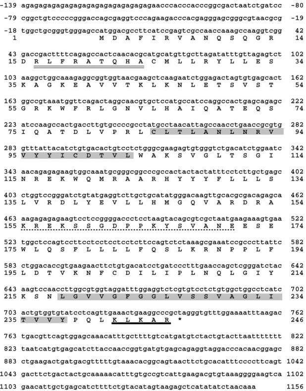
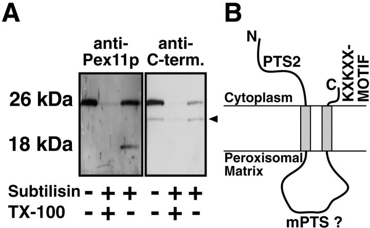
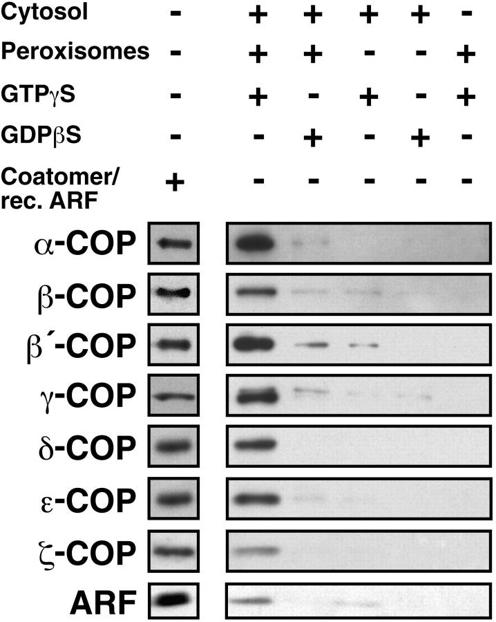
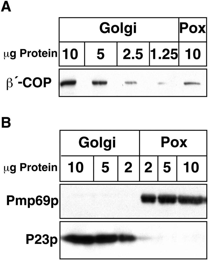
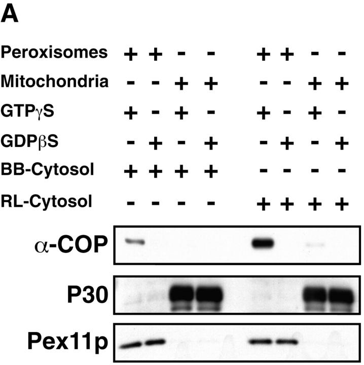
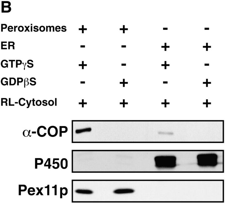
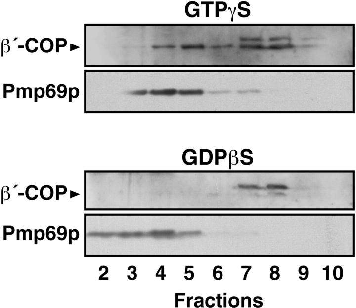
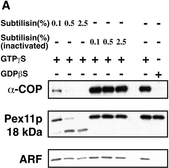
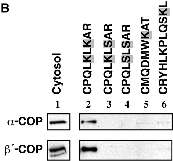
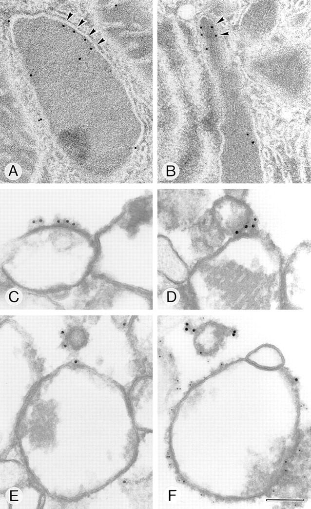
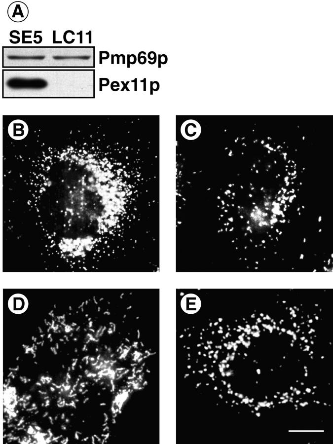
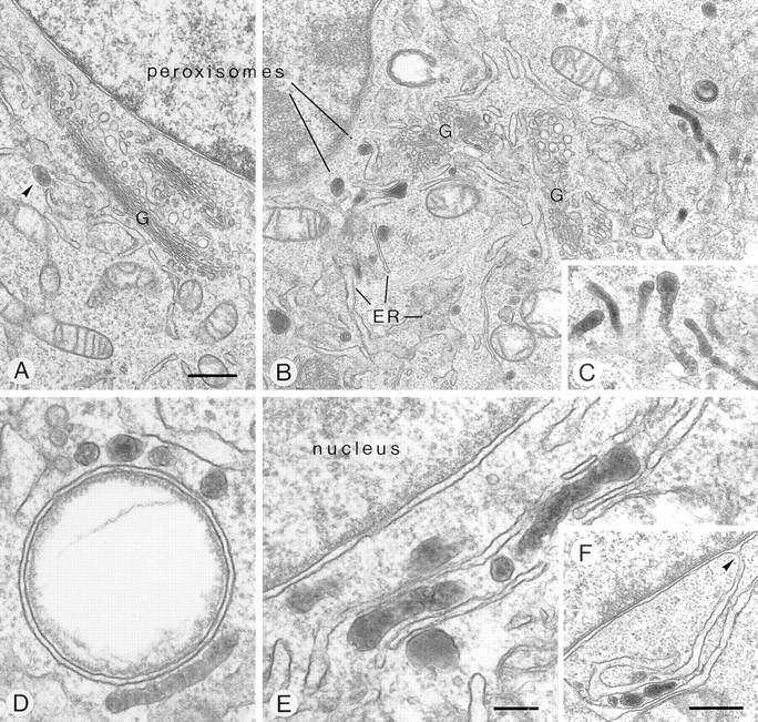
Peroxisomal membrane protein (Pmp)26p (RnPex11p), a major constituent of induced rat liver peroxisomal membrane, was found to contain a COOH-terminal, cytoplasmically exposed consensus dilysine motif with the potential to bind coatomer. Biochemical as well as immunocytochemical evidence is presented showing that peroxisomes incubated with preparations of bovine brain or rat liver cytosol recruit ADP-ribosylation factor (ARF) and coatomer in a strictly guanosine 5'-O-(3-thiotriphosphate)-dependent manner. Consistent with this observation, ldlF cells expressing a temperature-sensitive mutant version of the epsilon-subunit of coatomer exhibit elongated tubular peroxisomes possibly due to impaired vesiculation at the nonpermissive temperature. Since overexpression of Pex11p in Chinese hamster ovary wild-type cells causes proliferation of peroxisomes, these data suggest that Pex11p plays an important role in peroxisome biogenesis by supporting ARF- and coatomer-dependent vesiculation of the organelles.


Figures

Figure 1
Figure 1
Nucleotide and nucleotide-derived amino acid sequence of rat liver Pex11p-cDNA. Two putative transmembrane spans, as revealed by the Kyte-Doolittle algorithm (33) and the ProteinPredict program (55) are shaded. The consensus PTS2 sequence and the consensus-like sequence of a mPTS are underlined by a double and a single broken line, respectively. The COOH-terminal KXKXX dilysine motif is underlined in bold. These sequence data are available from GenBank/EMBL/DDBJ under accession number AJ224120.
Figure 2
Figure 2
Membrane topology of Pex11p. (A) Treatment of intact isolated rat liver peroxisomes (200 μg) with subtilisin (1 μg) shortened Pex11p to a 18-kD fragment that is recognized by the anti-Pex11p antiserum raised against the entire Pex11p (anti-Pex11p) but not by an antibody directed against its COOH-terminal peptide (anti-C-term.). The characteristic fragmentation of Pex11p is only obtained with intact peroxisomes and does not occur in the presence of Triton X-100 (TX-100). The antipeptide antiserum cross-reacted with an unidentified 23-kD peroxisomal polypeptide (arrowhead). (B) Proposed membrane topology of Pex11p. The polypeptide is inserted into the peroxisomal membrane by two hydrophobic transmembrane spans exposing the NH2 and COOH termini to the cytoplasm. mPTS, peroxisomal membrane targeting signal.
Figure 3
Figure 3
GTP-γS–dependent binding of coatomer and ARF to isolated rat liver peroxisomes. Peroxisomes (250 μg) were incubated with bovine brain cytosol (20 mg) in the presence of GTP-γS or GDP-βS (50 μM). Controls were run without peroxisomes and/or without cytosol. After incubation the organelles were recovered by centrifugation and 10-μg aliquots were subjected to SDS-PAGE and immunoblotting using monospecific antibodies against the seven coatomer subunits and ARF 1. The immunoreactivities of the different anti-COP antibodies against isolated bovine brain coatomer and recombinant ARF 1 are shown on the separate lane.
Figure 4
Figure 4
Analysis of Golgi contamination in isolated rat liver peroxisomal fractions. (A) Indicated amounts of isolated Golgi membranes and peroxisomes (Pox) primed with bovine brain cytosol in the presence of GTP-γS were subjected to SDS-PAGE and immunoblotting using an anti–β′-COP antibody. Note that the signal obtained with 10 μg of peroxisomes equals that of ∼2.5 μg of Golgi membranes. In B varying amounts of unprimed Golgi membranes and peroxisomes (Pox) were separated by SDS-PAGE and visualized by immunoblotting using antisera directed against Golgi P23 and Pmp69p, respectively. Note that the anti-P23 antiserum reliably recognizes 2 μg and even less of Golgi membranes and that even with 10 μg of peroxisomes no P23 immunoreaction product is observed.
Figure 5
Figure 5
Comparison of the coatomer binding activity of isolated rat liver peroxisomes, mitochondria, and microsomes. Peroxisomes or mitochondria (250 μg) were incubated with bovine brain (BB) or rat liver (RL) cytosol (10 mg) in the presence of GTP-γS or GDP-βS (50 μM). Coatomer binding was analyzed in aliquots of the reisolated organelles separated on SDS-PAGE by Western blotting using anti–α-COP antiserum. Peroxisomes and mitochondria were detected by anti-Pex11p and anti-P30 (30-kD inner mitochondrial membrane proteins) antiserum, respectively. Densitometric evaluation of the α-COP signal revealed that peroxisomes bind eight times more α-COP from rat liver than from bovine brain cytosol. Note that isolated mitochondria do not bind coatomer. (B) Peroxisomes and microsomes (250 μg) were incubated with rat liver cytosol (10 mg) in the presence of GTP-γS and GDP-βS (50 μM) and aliquots of the organelles, recovered by flotation, were analyzed for coatomer binding as above. Densitometric evaluation revealed that microsomes, detected by a polyclonal anti-P450 antiserum, bind coatomer six times less efficiently than peroxisomes.
Figure 5
Figure 5
Comparison of the coatomer binding activity of isolated rat liver peroxisomes, mitochondria, and microsomes. Peroxisomes or mitochondria (250 μg) were incubated with bovine brain (BB) or rat liver (RL) cytosol (10 mg) in the presence of GTP-γS or GDP-βS (50 μM). Coatomer binding was analyzed in aliquots of the reisolated organelles separated on SDS-PAGE by Western blotting using anti–α-COP antiserum. Peroxisomes and mitochondria were detected by anti-Pex11p and anti-P30 (30-kD inner mitochondrial membrane proteins) antiserum, respectively. Densitometric evaluation of the α-COP signal revealed that peroxisomes bind eight times more α-COP from rat liver than from bovine brain cytosol. Note that isolated mitochondria do not bind coatomer. (B) Peroxisomes and microsomes (250 μg) were incubated with rat liver cytosol (10 mg) in the presence of GTP-γS and GDP-βS (50 μM) and aliquots of the organelles, recovered by flotation, were analyzed for coatomer binding as above. Densitometric evaluation revealed that microsomes, detected by a polyclonal anti-P450 antiserum, bind coatomer six times less efficiently than peroxisomes.
Figure 6
Figure 6
GTP-γS–dependent colocalization of coatomer and peroxisomes in SLO-permeabilized hepatocytes after density gradient fractionation. Permeabilized hepatocytes were incubated with rat liver cytosol and GTP-γS or GDP-βS for 30 min at 37°C. After homogenization of the recovered cells the postnuclear supernate was fractionated on a linear Nycodenz density gradient. The distribution of coatomer and peroxisomes was visualized by Western blotting using rabbit anti–β′-COP and anti-Pmp69p peptide antisera, respectively. Note that the anti–β′-COP antiserum in the Golgi fractions (fractions 7 and 8) recognizes a cross-reaction product of molecular weight slightly higher than β′-COP.
Figure 7
Figure 7
Involvement of the dilysine motif of Pex11p in peroxisomal coatomer binding. (A) Binding of coatomer and ARF to peroxisomes (250 μg) pretreated with increasing concentrations of subtilisin, as indicated, was performed by incubations with rat liver cytosol (10 mg) in the presence of GTP-γS or GDP-βS (50 μM). Controls remained untreated or were treated with the same concentrations of subtilisin previously inactivated by 1 mM PMSF. Peroxisomes were identified by the polyclonal anti-Pex11p antiserum that recognizes both the uncleaved Pex11p as well as the 18-kD proteolytic fragment (see legend to Fig. 2), whereas coatomer and ARF binding was detected by anti– α-COP and anti-ARF 1 antiserum, respectively. Note that proteolytic degradation of Pex11p correlates with a decrease in binding of both α-COP and ARF. (B) Selective binding of coatomer to the COOH-terminal Pex11p tail peptide containing the dilysine motif. The synthetic peptide (lane 2) and two mutated versions of it (lanes 3 and 4) as well as the COOH-terminal octapeptide of ScPmp27p (lane 5) and an undecapeptide containing the nine COOH-terminal amino acid residues of rat peroxisomal acyl-CoA oxidase (lane 6) were coupled to thiopropyl–Sepharose via an NH2 terminally attached cysteine residue. After incubation with bovine brain cytosol and extensive washing of the Sepharose, bound coatomer was released by SDS and visualized by SDS-PAGE and immunoblotting using monospecific antisera directed against α- and β′-COP. An aliquot (50 μg) of cytosol used in these incubations was loaded onto the gel as a control (lane 1).
Figure 7
Figure 7
Involvement of the dilysine motif of Pex11p in peroxisomal coatomer binding. (A) Binding of coatomer and ARF to peroxisomes (250 μg) pretreated with increasing concentrations of subtilisin, as indicated, was performed by incubations with rat liver cytosol (10 mg) in the presence of GTP-γS or GDP-βS (50 μM). Controls remained untreated or were treated with the same concentrations of subtilisin previously inactivated by 1 mM PMSF. Peroxisomes were identified by the polyclonal anti-Pex11p antiserum that recognizes both the uncleaved Pex11p as well as the 18-kD proteolytic fragment (see legend to Fig. 2), whereas coatomer and ARF binding was detected by anti– α-COP and anti-ARF 1 antiserum, respectively. Note that proteolytic degradation of Pex11p correlates with a decrease in binding of both α-COP and ARF. (B) Selective binding of coatomer to the COOH-terminal Pex11p tail peptide containing the dilysine motif. The synthetic peptide (lane 2) and two mutated versions of it (lanes 3 and 4) as well as the COOH-terminal octapeptide of ScPmp27p (lane 5) and an undecapeptide containing the nine COOH-terminal amino acid residues of rat peroxisomal acyl-CoA oxidase (lane 6) were coupled to thiopropyl–Sepharose via an NH2 terminally attached cysteine residue. After incubation with bovine brain cytosol and extensive washing of the Sepharose, bound coatomer was released by SDS and visualized by SDS-PAGE and immunoblotting using monospecific antisera directed against α- and β′-COP. An aliquot (50 μg) of cytosol used in these incubations was loaded onto the gel as a control (lane 1).
Figure 8
Figure 8
Peroxisomal localization of Pex11p (A and B) and coatomer binding (C–F) by immunogold electron microscopy (post- [A and B] and preembedding [C–F]). Clusters of Pex11p (arrowheads) are localized to the peroxisomal membrane in rat liver treated with clofibrate (A) or trifluoroacetate (B). Coatomer binding to clofibrate-induced isolated rat liver peroxisomes primed with bovine brain cytosol and GTP-γS was detected on peroxisomes, buds, and small vesicular structures. Note the nucleoid core in D and E that clearly distinguishes these organelles as peroxisomes. The mouse monoclonal antibody CM1A10 decorated with 12-nm gold-labeled second antibody was used to detect coatomer (C–F). A polyclonal anti-Pex11p tail peptide antiserum decorated with 14-nm protein A–gold (A and B) and anti-Pmp69p peptide antiserum decorated with 6-nm gold-labeled second antibody were used to detect the peroxisomal membrane (F). Bar, 100 nm.
Figure 9
Figure 9
Phenotypic change in peroxisomal morphology by overexpression of Pex11p (A–C) as well as functional deficiency of coatomer (D, E). CHO cells were stably transfected with either Pex11p-cDNA cloned into pcDNA3 (SE5) or pcDNA3 lacking an insert (LC11). Carbonate membranes of postnuclear supernates (10 μg) were analyzed for their content of Pmp69p and Pex11p by SDS-PAGE and Western blotting (A). The peroxisomal compartment of SE5 (B) and LC11 (C) was visualized by immunofluorescence using the polyclonal anti-Pmp69p antiserum. ldlF cells expressing a ts mutant of ε-COP were kept for 24 h at nonpermissive (39.5°C, D) or permissive (34°C, E) temperature before immunofluorescence staining of peroxisomes using the anti-Pmp69p antiserum. Note numerical increase in small spherical peroxisomes (B) and clustering of tubular peroxisomes (D). Bar, 3 μm.
Figure 10
Figure 10
Fine structure of peroxisomes in the perinuclear region of mutant ldlF cells at permissive (34°C, A, arrowhead) and nonpermissive (39.5°C, B–F) temperature. Phenotypic peroxisome clustering and formation of peroxisome-ER aggregates is revealed, due to functional deficiency of ε-COP. At nonpermissive temperature, the intensely DAB-stained peroxisomes frequently exhibit a highly tortuous, tubular shape (B–E). They cluster at the periphery of lipid vacuoles (D) and form rows alternating with ER cisternae (E), the latter of which are often in continuity with the perinuclear membrane (F, arrowhead). G, Golgi complex. Bars: (A–C and F) 500 nm; (D and E) 150 nm.

References

    1. Aniento F, Gu F, Parton RG, Gruenberg J. An endosomal β-COP is involved in the pH-dependent formation of transport vesicles destined for late endosomes. J Cell Biol. 1996;133:29–41. - PMC - PubMed
    1. Antoine B, Levrat F, Vallet V, Berbar T, Cartier N, Dubois N, Briand P, Kahn A. Gene expression in hepatocyte-like lines established by targeted carcinogenesis in transgenic mice. Exp Cell Res. 1992;200:175–185. - PubMed
    1. Aridor M, Bannykh SI, Rowe T, Balch WE. Sequential coupling between COPII and COPI vesicle coats in endoplasmic reticulum to Golgi transport. JCell Biol. 1995;131:875–893. - PMC - PubMed
    1. Baerends RJ, Rasmussen SW, Hilbrands RE, van der Heide M, Faber KN, Reuvekamp PT, Kiel JA, Cregg JM, van der Klei IJ, Veenhuis M. The Hansenula polymorphaPER9 gene encodes a peroxisomal membrane protein essential for peroxisome assembly and integrity. J Biol Chem. 1996;271:8887–8894. - PubMed
    1. Barlowe C, Orci L, Yeung T, Hosobuchi M, Hamamoto S, Salama N, Rexach MF, Ravazzola M, Amherdt M, Schekman R. COPII: a membrane coat formed by Sec proteins that drive vesicle budding from the endoplasmic reticulum. Cell. 1994;77:895–908. - PubMed

Publication types

MeSH terms

Associated data