. 1998 Apr 20;141(2):397-408.
doi: 10.1083/jcb.141.2.397.

Occludin-deficient embryonic stem cells can differentiate into polarized epithelial cells bearing tight junctions

Affiliations

Occludin-deficient embryonic stem cells can differentiate into polarized epithelial cells bearing tight junctions

M Saitou et al. J Cell Biol. .

Abstract

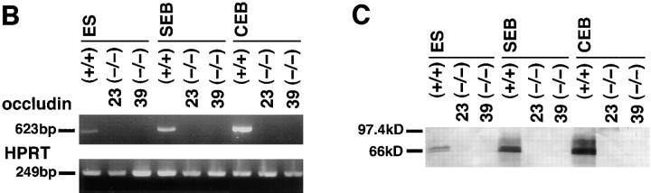
Occludin is the only known integral membrane protein of tight junctions (TJs), and is now believed to be directly involved in the barrier and fence functions of TJs. Occludin-deficient embryonic stem (ES) cells were generated by targeted disruption of both alleles of the occludin gene. When these cells were subjected to suspension culture, they aggregated to form simple, and then cystic embryoid bodies (EBs) with the same time course as EB formation from wild-type ES cells. Immunofluorescence microscopy and ultrathin section electron microscopy revealed that polarized epithelial (visceral endoderm-like) cells were differentiated to delineate EBs not only from wild-type but also from occludin-deficient ES cells. Freeze fracture analyses indicated no significant differences in number or morphology of TJ strands between wild-type and occludin-deficient epithelial cells. Furthermore, zonula occludens (ZO)-1, a TJ-associated peripheral membrane protein, was still exclusively concentrated at TJ in occludin-deficient epithelial cells. In good agreement with these morphological observations, TJ in occludin-deficient epithelial cells functioned as a primary barrier to the diffusion of a low molecular mass tracer through the paracellular pathway. These findings indicate that there are as yet unidentified TJ integral membrane protein(s) which can form strand structures, recruit ZO-1, and function as a barrier without occludin.


Figures

Figure 1
Figure 1
Targeted inactivation of the occludin gene in ES cells. (A) Restriction maps of the wild-type allele, the targeting vector, and the targeted allele of the occludin gene. The first ATG codon was located in exon 2; exon 3 encoded the NH2-terminal half of occludin molecule from the first transmembrane domain to the second extracellular loop. Exon 4 encoded the fourth transmembrane domain and the initial part of the COOH-terminal cytoplasmic domain, indicating the existence of one or more downstream exons. The targeting vector contained the SA IRES/ LacZ/loxP/pgk neo/loxP cassette in its middle portion to delete exon 3 in the targeted allele. The positions of 5′, 3′, and neo probes for Southern blotting are indicated as bars. The loxP sequence flanking pgk neo is shown by closed triangles. P, PstI; X, XbaI; N, NcoI; E, EcoRV. (B) Generation of occludin single knock-out ES cell lines. Southern blotting analysis of XbaI- digested occludin locus with 3′ probe (3probe) yielded a 9.2-kb band from the wild-type allele (clone 464) and a 4.6-kb band from the targeted allele (clone 380). The correct integration of the vector was confirmed by Southern blotting of PstI/EcoRV digest with 5′ probe (5probe). The 15.5- and 5.4-kb bands were derived from wild-type and targeted alleles, respectively. Furthermore, targeted clones were checked for single integration by hybridization with a neo probe (neo probe). (C) Generation of occludin double knock-out ES cell lines. Two independent double knock-out clones (clones 23 and 39) were obtained from the single knock-out clone 380 in the presence of an elevated concentration of G418. The double knock-out cells lost the normal 9.2-kb band, indicating complete knock-out. Theoretically, it is possible that deleted COOH-terminal fragments of occludin (encoded by exons 4, 5 ---) are expressed in these cells, but these fragments, if any, are not regarded as structural or functional components of TJ strands.
Figure 2
Figure 2
Embryoid body formation and occludin expression. (A) The appearance of ES cells (a and e), simple embryoid bodies (EBs) (b and f), and cystic EBs (c, d, g, and h) bearing homozygous wild-type (a–d) or targeted (e–h) occludin genes. No differences were detected between wild-type and occludin double knock-out ES cells/EBs, not only in their morphological appearance but also in the time course of EB formation. Regardless of occludin genotype, ES cells were grown on feeder cells with undifferentiated morphology (a and e). Simple (b and f) and cystic EBs (c and g) were formed in 4- and 10-d suspension culture, respectively. Simple EBs were characterized by Reichert's membranes (arrowheads), suggesting differentiation of the outermost visceral endoderm-like cell layer. Cystic EBs were fully expanded with internally secreted liquid (c and g), which was also shown on toluidine blue–stained semi-thin sectional images (d and h). (B) Loss of occludin mRNA in occludin double knock-out ES cells/EBs examined by RT-PCR. A trace amount of occludin mRNA was detected in wild-type ES cells (ES), and the amount appeared to increase as ES cells differentiated into simple EBs (SEB) and then into cystic EBs (CEB). In contrast, occludin expression was completely abolished in occludin double knock-out ES cells (clones 23 and 39), simple, and cystic EBs. As a control, the hypoxanthine phosphoribosyl transferase gene was equally amplified in all samples. (C) Immunoblotting analyses with anti-occludin pAb. Consistent with the RT-PCR data, the protein expression level of occludin was elevated during the course of wild-type EB development, whereas no occludin expression was detected in occludin double knock-out ES cells/EBs. Bars: (a, b, e, and f) 100 μm; (c and g) 500 μm; (d and h) 200 μm.
Figure 2
Figure 2
Embryoid body formation and occludin expression. (A) The appearance of ES cells (a and e), simple embryoid bodies (EBs) (b and f), and cystic EBs (c, d, g, and h) bearing homozygous wild-type (a–d) or targeted (e–h) occludin genes. No differences were detected between wild-type and occludin double knock-out ES cells/EBs, not only in their morphological appearance but also in the time course of EB formation. Regardless of occludin genotype, ES cells were grown on feeder cells with undifferentiated morphology (a and e). Simple (b and f) and cystic EBs (c and g) were formed in 4- and 10-d suspension culture, respectively. Simple EBs were characterized by Reichert's membranes (arrowheads), suggesting differentiation of the outermost visceral endoderm-like cell layer. Cystic EBs were fully expanded with internally secreted liquid (c and g), which was also shown on toluidine blue–stained semi-thin sectional images (d and h). (B) Loss of occludin mRNA in occludin double knock-out ES cells/EBs examined by RT-PCR. A trace amount of occludin mRNA was detected in wild-type ES cells (ES), and the amount appeared to increase as ES cells differentiated into simple EBs (SEB) and then into cystic EBs (CEB). In contrast, occludin expression was completely abolished in occludin double knock-out ES cells (clones 23 and 39), simple, and cystic EBs. As a control, the hypoxanthine phosphoribosyl transferase gene was equally amplified in all samples. (C) Immunoblotting analyses with anti-occludin pAb. Consistent with the RT-PCR data, the protein expression level of occludin was elevated during the course of wild-type EB development, whereas no occludin expression was detected in occludin double knock-out ES cells/EBs. Bars: (a, b, e, and f) 100 μm; (c and g) 500 μm; (d and h) 200 μm.
Figure 3
Figure 3
Immunofluorescence confocal microscopic localization of occludin and ZO-1 in wild-type and occludin-deficient simple EBs (a–f)/ cystic EBs (g–l). Frozen sections of EBs were doubly stained with rat anti-occludin mAb (a, d, g, and j) and mouse anti–ZO-1 mAb (b, e, h, and k). In both wild-type simple (a–c) and cystic (g–i) EBs, occludin was specifically expressed in the outermost cell layers, and precisely colocalized with ZO-1 probably at junctional regions. In contrast, in occludin-deficient simple (d–f) and cystic (j–l) EBs, occludin disappeared from the outermost cell layers (d and j), and the loss of occludin expression did not appear to affect the distribution of ZO-1 (e and k). Bar, 10 μm.
Figure 6
Figure 6
Subcellular distribution of ZO-1 in occludin-deficient epithelial cells. (A) Immunofluorescence confocal microscopy. Frozen sections of wild-type (a–f) and occludin-deficient (g–i) cystic EBs were doubly labeled with the mixture of rat anti-occludin mAb (a) and mouse anti–ZO-1 mAb (b) or the mixture of rat anti–E-cadherin mAb (d and g) and mouse anti–ZO-1 mAb (e and h). In wild-type EBs, E-cadherin was distributed along lateral membranes as well as at junctional regions (d), whereas occludin (a) and ZO-1 (b and e) were highly concentrated at junctional regions. Close comparison revealed that at junctional regions ZO-1 was precisely colocalized with occludin (c), but located more apically than E-cadherin (f). The same spatial relationship between E-cadherin (g) and ZO-1 (h) was maintained in occludin-deficient EBs (i). (B) Immunoelectron microscopy. Ultrathin cryosections of occludin-deficient cystic EBs were labeled with mouse anti–ZO-1 mAb. TJ was exclusively labeled. Bars: (A) 10 μm; (B) 200 nm.
Figure 6
Figure 6
Subcellular distribution of ZO-1 in occludin-deficient epithelial cells. (A) Immunofluorescence confocal microscopy. Frozen sections of wild-type (a–f) and occludin-deficient (g–i) cystic EBs were doubly labeled with the mixture of rat anti-occludin mAb (a) and mouse anti–ZO-1 mAb (b) or the mixture of rat anti–E-cadherin mAb (d and g) and mouse anti–ZO-1 mAb (e and h). In wild-type EBs, E-cadherin was distributed along lateral membranes as well as at junctional regions (d), whereas occludin (a) and ZO-1 (b and e) were highly concentrated at junctional regions. Close comparison revealed that at junctional regions ZO-1 was precisely colocalized with occludin (c), but located more apically than E-cadherin (f). The same spatial relationship between E-cadherin (g) and ZO-1 (h) was maintained in occludin-deficient EBs (i). (B) Immunoelectron microscopy. Ultrathin cryosections of occludin-deficient cystic EBs were labeled with mouse anti–ZO-1 mAb. TJ was exclusively labeled. Bars: (A) 10 μm; (B) 200 nm.
Figure 4
Figure 4
Ultrathin section electron microscopy images of outermost cell layers of wild-type (a, c, and e) and occludin-deficient EBs (b, d, and f). Both in wild-type and occludin-deficient simple EBs (a and b), an epithelial cell layer was differentiated with microvilli-associated apical membrane domains. Also, in both wild-type and occludin-deficient cystic EBs (c–f), the epithelial cells were further polarized with numerous microvilli (arrows) and secretory granules (asterisks). At the most apical region of the lateral membranes of these epithelial cells, well-developed tight junctions (TJ) were easily identified in wild-type (e) as well as occludin-deficient EBs (f). DS, desmosome. Bars: (a and b) 2 μm; (c and d) 7 μm; (e and f) 200 nm.
Figure 4
Figure 4
Ultrathin section electron microscopy images of outermost cell layers of wild-type (a, c, and e) and occludin-deficient EBs (b, d, and f). Both in wild-type and occludin-deficient simple EBs (a and b), an epithelial cell layer was differentiated with microvilli-associated apical membrane domains. Also, in both wild-type and occludin-deficient cystic EBs (c–f), the epithelial cells were further polarized with numerous microvilli (arrows) and secretory granules (asterisks). At the most apical region of the lateral membranes of these epithelial cells, well-developed tight junctions (TJ) were easily identified in wild-type (e) as well as occludin-deficient EBs (f). DS, desmosome. Bars: (a and b) 2 μm; (c and d) 7 μm; (e and f) 200 nm.
Figure 5
Figure 5
Freeze fracture images of tight junctions in the outermost epithelial cell layers of wild-type (a, c, e, f, and i) and occludin-deficient cystic EBs (b, d, g, h, and j). In both wild-type and occludin-deficient EBs, well-developed continuous and anastomosing TJ strands were detected. Although the number of TJ strands significantly varied from cell to cell and from EB to EB, it was distributed within the similar range regardless of occludin expression; in wild-type and occludin-deficient EBs it fell within the range from a to c and from b to d, respectively. P-face TJ strands (e and g) and complementary E-face TJ grooves (f and h) were also indistinguishable in their morphology between wild-type and occludin-deficient EBs. By immunoreplica electron microscopy, TJ strands of wild-type EBs (i) were heavily labeled with anti-occludin mAb, whereas those from occludin-deficient EBs (j) were not labeled. Bars: (a–d) 200 nm; (e–h) 50 nm; (i–j) 100 nm.
Figure 5
Figure 5
Freeze fracture images of tight junctions in the outermost epithelial cell layers of wild-type (a, c, e, f, and i) and occludin-deficient cystic EBs (b, d, g, h, and j). In both wild-type and occludin-deficient EBs, well-developed continuous and anastomosing TJ strands were detected. Although the number of TJ strands significantly varied from cell to cell and from EB to EB, it was distributed within the similar range regardless of occludin expression; in wild-type and occludin-deficient EBs it fell within the range from a to c and from b to d, respectively. P-face TJ strands (e and g) and complementary E-face TJ grooves (f and h) were also indistinguishable in their morphology between wild-type and occludin-deficient EBs. By immunoreplica electron microscopy, TJ strands of wild-type EBs (i) were heavily labeled with anti-occludin mAb, whereas those from occludin-deficient EBs (j) were not labeled. Bars: (a–d) 200 nm; (e–h) 50 nm; (i–j) 100 nm.
Figure 7
Figure 7
Tight junction permeability assay of wild-type and occludin-deficient epithelial cells. After wild-type (a and b) or occludin-deficient (e and f) cystic EBs were incubated with NHS-LC-biotin for 30 min, frozen sections were incubated with XRITC-avidin to localize surface-bound biotin (a and e). To visualize the contours of each epithelial cell, frozen sections were counterstained with fluorescein-phalloidin (b, d, and f). Regardless of occludin expression, only the apical surface of the outermost epithelial cell layer was biotinylated, indicating no paracellular leakage of NHS-LC-biotin. In contrast, when wild-type EBs were incubated with NHS-LC-biotin in the presence of 10 mM EGTA to affect intercellular junctions, the basolateral membranes as well as the apical membranes of epithelial cells were equally biotinylated (c). Bar, 10 μm.

References

    1. Anderson JM, Fanning AS, Lapierre L, Van Itallie CM. Zonula occludens (ZO)-1 and ZO-2: membrane-associated guanylate kinase homologues (MAGuKs) of the tight junction. Biochem Soc Trans. 1995;23:470–475. - PubMed
    1. Ando-Akatsuka Y, Saitou M, Hirase T, Kishi M, Sakakibara A, Itoh M, Yonemura S, Furuse M, Sh, Tsukita Interspecies diversity of the occludin sequence: cDNA cloning of human, mouse, dog, and rat-kangaroo homologues. J Cell Biol. 1996;133:43–47. - PMC - PubMed
    1. Anzini P, Neuberg DH-H, Schachner M, Nelles E, Willecke K, Zielasek J, Toyka KV, Suter U, Martini R. Structural abnormalities and deficient maintenance of peripheral nerve myelin in mice lacking the gap junction protein connexin 32. J Neurosci. 1997;17:4545–4551. - PMC - PubMed
    1. Balda MS, Whitney JA, Flores C, González S, Cereijido M, Matter K. Functional dissociation of paracellular permeability and transepithelial electrical resistance and disruption of the apical-basolateral intramembrane diffusion barrier by expression of a mutant tight junction membrane protein. J Cell Biol. 1996;134:1031–1049. - PMC - PubMed
    1. Baribault H, Oshima RG. Polarized and functional epithelia can form after the targeted inactivation of both mouse keratin 8 alleles. J Cell Biol. 1991;115:1675–1684. - PMC - PubMed

Publication types

MeSH terms