. 1998 Apr 20;141(2):503-14.
doi: 10.1083/jcb.141.2.503.

p21 is a critical CDK2 regulator essential for proliferation control in Rb-deficient cells

Affiliations

p21 is a critical CDK2 regulator essential for proliferation control in Rb-deficient cells

J Brugarolas et al. J Cell Biol. .

Abstract

Proliferation in mammalian cells is controlled primarily in the G1-phase of the cell cycle through the action of the G1 cyclin-dependent kinases, CDK4 and CDK2. To explore the mechanism of cellular response to extrinsic factors, specific loss of function mutations were generated in two negative regulators of G1 progression, p21 and pRB. Individually, these mutations were shown to have significant effects in G1 regulation, and when combined, Rb and p21 mutations caused more profound defects in G1. Moreover, cells deficient for pRB and p21 were uniquely capable of anchorage-independent growth. In contrast, combined absence of pRB and p21 function was not sufficient to overcome contact inhibition of growth nor for tumor formation in nude mice. Finally, animals with the genotype Rb+/-;p21(-/-) succumbed to tumors more rapidly than Rb+/- mice, suggesting that in certain contexts mutations in these two cell cycle regulators can cooperate in tumor development.


Figures

Figure 1
Figure 1
CDK2 and CDK4 catalytic activities from asynchronously growing wild-type (Wt), p21 −/−, Rb −/−, and Rb −/−;p21 −/− cells. (A) In vitro CDK4 kinase assay using GST-RB as a substrate. (B) In vitro CDK2 kinase assay using GST-RB as a substrate. (C) Histogram representing quantitative analysis of the results from the CDK2 kinase assay. (D) Western blot analysis of p21 and pRB in wild-type, p21 −/−, and Rb −/− cells. Lanes shown were reassembled in appropriate order from a single autoradiogram. All samples were derived from the same experiment.
Figure 2
Figure 2
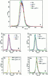
Cell size analysis of wild-type, p21 −/−, Rb −/−, and Rb −/−; p21 −/− MEFs. Asynchronously growing cells were harvested, fixed, stained with PI and analyzed by FACS®. 60,000 events were collected for each sample. 2n and 4n DNA containing populations were gated for cell size analysis in a FSC-H by FL2-A dot plot and scored in a FSC-H histogram. (A) Cell size analysis of wild-type, p21 −/−, Rb −/−, and Rb −/−;p21 −/− cells. (B and C) Cell size analysis of wild-type, p21 −/−, Rb −/−, and Rb −/−;p21 −/− cells with a 2n DNA content. (D) Size analysis of two strains of Rb −/−; p21 −/− cells with a 2n DNA content. These strains were derived from two different embryos. (E) Cell size analysis of wild-type, p21 −/−, Rb −/−, and Rb −/−;p21 −/− cells with a 4n DNA content.
Figure 3
Figure 3
Cell cycle analysis of G0-S phase interval in Rb −/−;p21 −/− and wild-type MEFs and growth rates of Rb −/−;p21 −/−, Rb −/−, p21 −/−, and wild-type cells. (A–F) DNA content profiles of synchronous cultures of wild-type and Rb −/−;p21 −/− cells at different times after release from G0. (G) Growth properties of Rb −/−;p21 −/−, Rb −/−, p21 −/−, and wild-type MEFs derived from littermate embryos in tissue culture media supplemented with 10% FCS. Values at different timepoints represent the average of the measurements from duplicate samples. This experiment was performed three times with duplicate samples with similar results.
Figure 4
Figure 4
Growth properties of wild-type, p21 −/−, Rb −/−, and Rb −/−;p21 −/− cells in the presence of limiting concentrations of growth factors. (A and B) Cells were synchronized in G0 and replated at low density in 0.1 or 0.5% FCS. At the indicated times, duplicate samples were harvested and counted. Similar results were obtained at 0.05 and 0.3% FCS (data not shown). (C) Comparative analysis of the growth properties of Rb −/−;p21 −/− cells in 0.5 and 5% FCS.
Figure 5
Figure 5
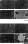
Anchorage-dependence for growth of wild-type, p21 −/−, Rb −/−, and Rb −/−;p21 −/− cells. G0 synchronized populations were plated in soft agar and colony formation was scored after 3 wk. Bars: (A–D and E–H) 500 μm.
Figure 6
Figure 6
Cyclin D1 and p27 protein levels, and CDK2 kinase activity in wild-type and Rb −/−;p21 −/− cells grown in monolayer (Mn.) and in suspension (Sp.). (A) Cyclin D1 protein levels assayed by Western blot analysis. (B) p27 protein levels assayed by Western blot analysis. (C) In vitro CDK2 kinase assay using GST-RB as a substrate. (D) Histogram representing quantitation analysis of the results from the CDK2 kinase assay. (E) In vitro CDK2 kinase assay of wild-type, p21 −/−, and Rb −/− ;p21 −/− cells in suspension using GST-RB as a substrate. Lanes shown were reassembled in appropriate order from a single autoradiogram. All samples were derived from the same experiment.
Figure 7
Figure 7
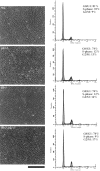
Sensitivity to contact inhibition of growth of wild-type, p21 −/−, Rb −/−, and Rb −/−;p21 −/− cells. The left column shows representative fields of confluent cultures. The right column shows cell cycle profiles of confluent cultures after 6 d. 10,000 events were collected for each sample. The data were analyzed using ModFit LT software (Becton Dickinson). The coefficient of variation was below 6 for all the samples. Bar, 500 μm.
Figure 8
Figure 8
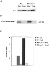
p27 protein levels and CDK2 kinase activity from exponentially growing (Exp.) and confluent (Conf.) wild-type, and Rb −/−;p21 −/− cells. (A) p27 protein levels analyzed by Western blot analysis. (B) In vitro CDK2 kinase assay using GST-RB as a substrate. (C) Histogram representing quantitative analysis of the results from the CDK2 kinase assay.
Figure 9
Figure 9
Survival curve of p21 −/−;Rb +/− mice (n = 70) compared with Rb +/− mice (n = 50; Williams et al., 1994) in a mixed C57BL/ 6-129/Sv genetic background.

References

    1. Assoian RK. Anchorage-dependent cell cycle progression. J Cell Biol. 1997;136:1–4. - PMC - PubMed
    1. Baldin V, Lukas J, Marcote MJ, Pagano M, Draetta J. Cyclin D1 is a nuclear protein required for progression in G1. Genes Dev. 1993;7:812–821. - PubMed
    1. Bohmer R, Scharf E, Assoian RK. Cytoskeletal integrity is required throughout the mitogen stimulation phase of the cell cycle and mediates anchorage-dependent expression of cyclin D1. Mol Biol Cell. 1996;7:101–111. - PMC - PubMed
    1. Brugarolas J, Chandrasekaran C, Gordon JI, Beach D, Jacks T, Hannon GJ. Radiation-induced cell cycle arrest compromised by p21 deficiency. Nature. 1995;377:552–557. - PubMed
    1. Chellappan SP, Hiebert S, Mudryj M, Horowitz JM, Nevins JR. The E2F transcription factor is a cellular target for the RB protein. Cell. 1991;65:1053–1061. - PubMed

MeSH terms