. 1998 Apr 20;141(2):539-51.
doi: 10.1083/jcb.141.2.539.

Rho-mediated contractility exposes a cryptic site in fibronectin and induces fibronectin matrix assembly

Affiliations

Rho-mediated contractility exposes a cryptic site in fibronectin and induces fibronectin matrix assembly

C Zhong et al. J Cell Biol. .

Abstract

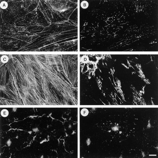
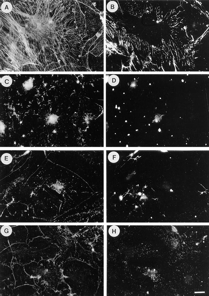
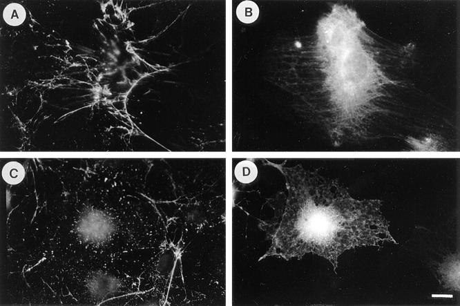
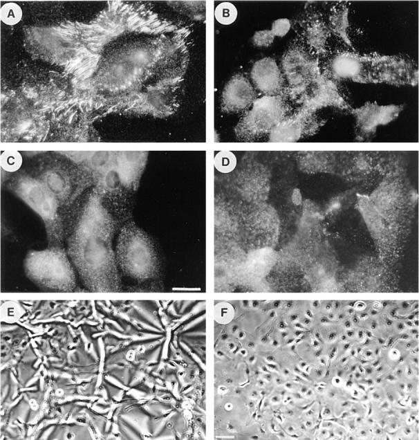
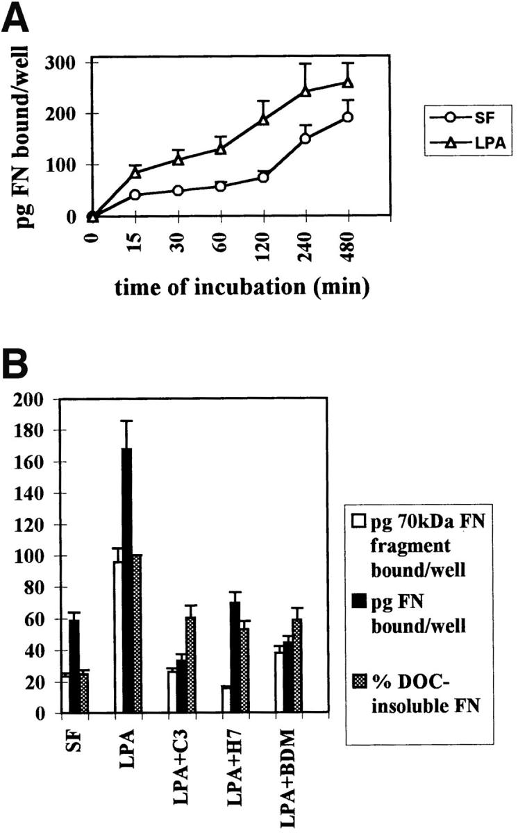
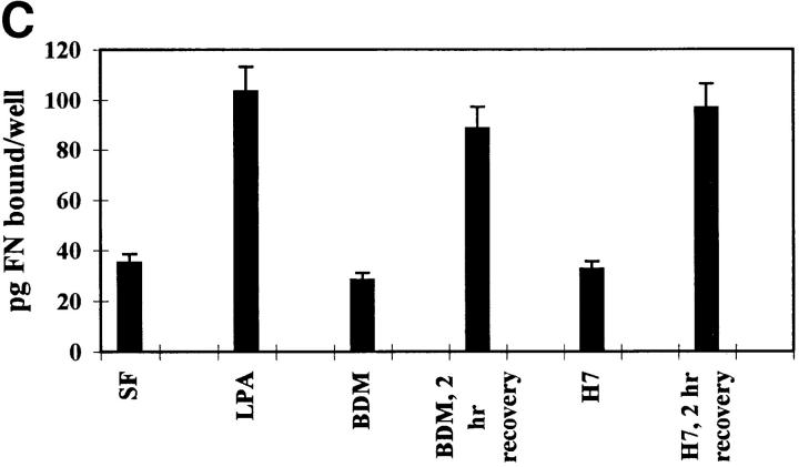
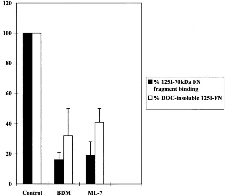
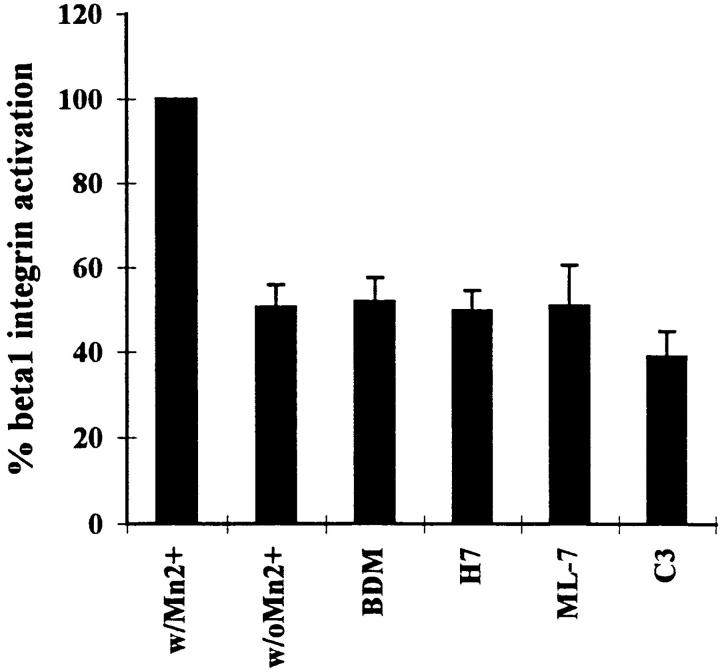
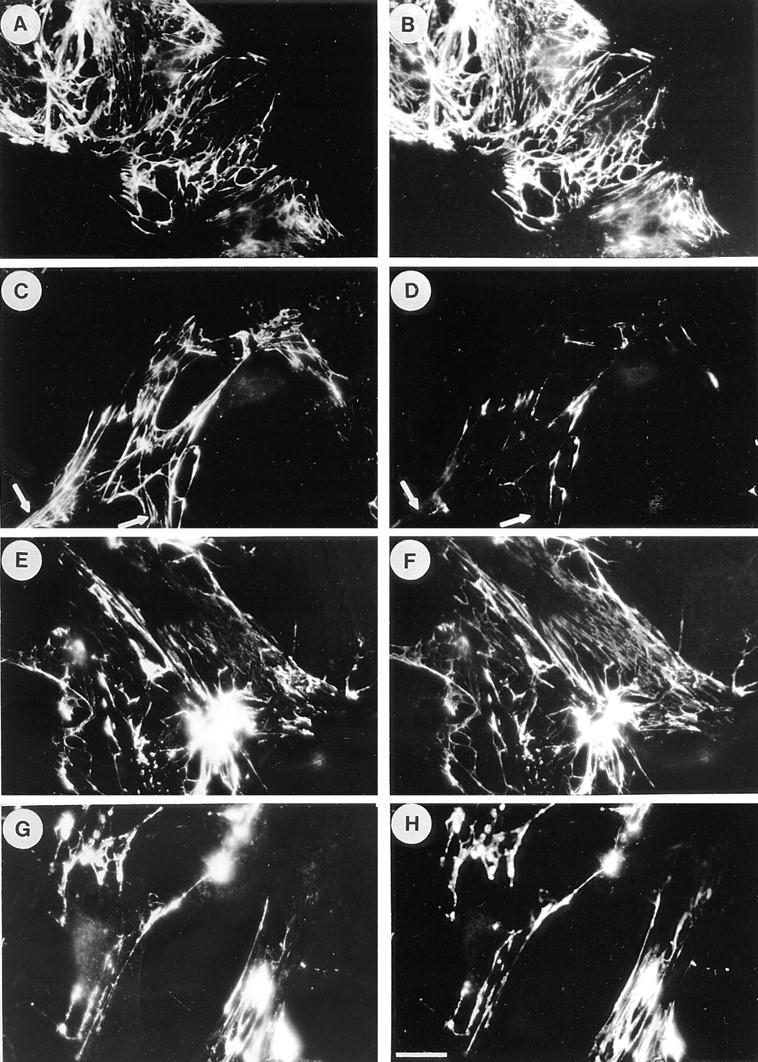
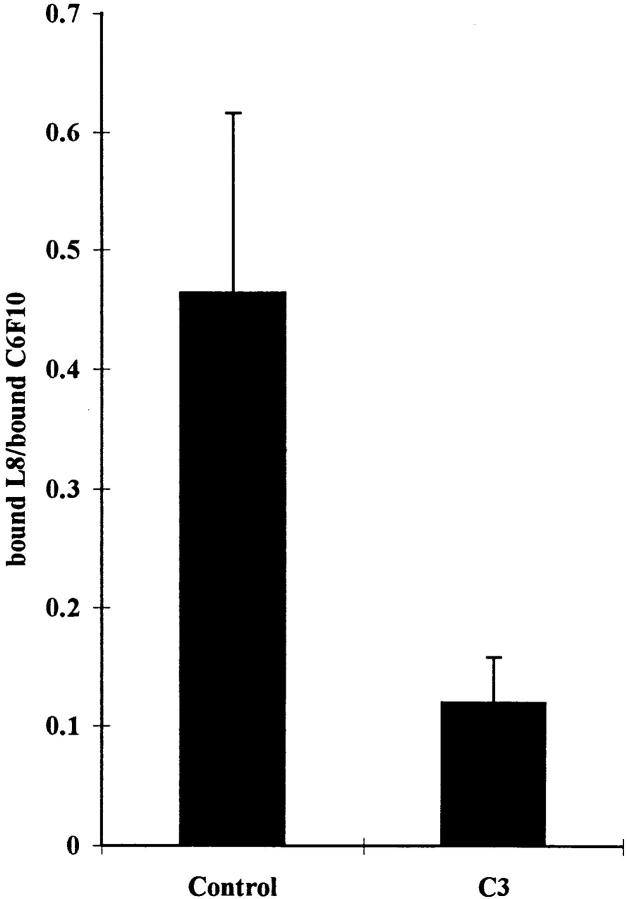
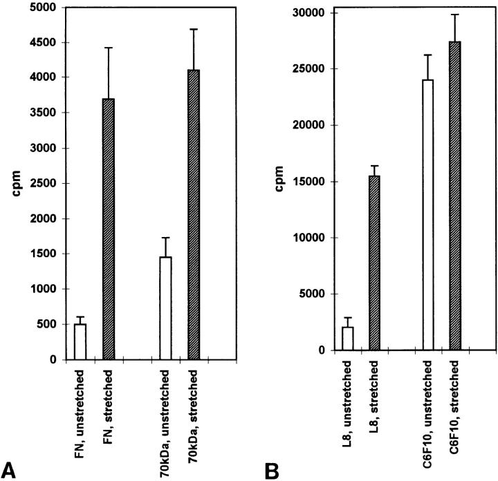
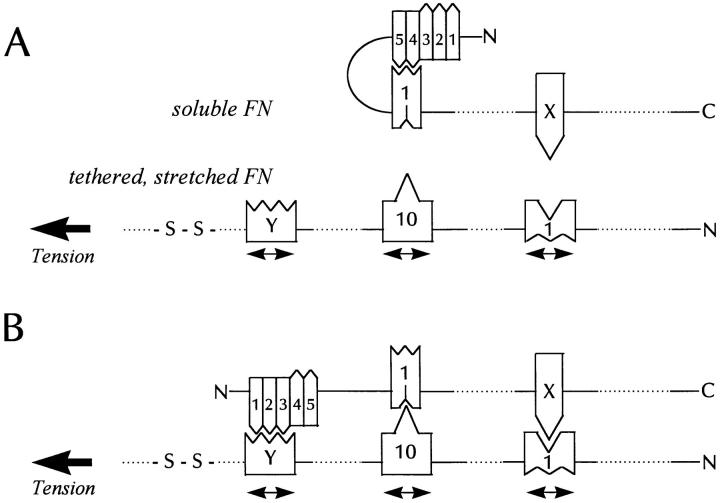
Many factors influence the assembly of fibronectin into an insoluble fibrillar extracellular matrix. Previous work demonstrated that one component in serum that promotes the assembly of fibronectin is lysophosphatidic acid (Zhang, Q., W.J. Checovich, D.M. Peters, R.M. Albrecht, and D.F. Mosher. 1994. J. Cell Biol. 127:1447-1459). Here we show that C3 transferase, an inhibitor of the low molecular weight GTP-binding protein Rho, blocks the binding of fibronectin and the 70-kD NH2-terminal fibronectin fragment to cells and blocks the assembly of fibronectin into matrix induced by serum or lysophosphatidic acid. Microinjection of recombinant, constitutively active Rho into quiescent Swiss 3T3 cells promotes fibronectin matrix assembly by the injected cells. Investigating the mechanism by which Rho promotes fibronectin polymerization, we have used C3 to determine whether integrin activation is involved. Under conditions where C3 decreases fibronectin assembly we have only detected small changes in the state of integrin activation. However, several inhibitors of cellular contractility, that differ in their mode of action, inhibit cell binding of fibronectin and the 70-kD NH2-terminal fibronectin fragment, decrease fibronectin incorporation into the deoxycholate insoluble matrix, and prevent fibronectin's assembly into fibrils on the cell surface. Because Rho stimulates contractility, these results suggest that Rho-mediated contractility promotes assembly of fibronectin into a fibrillar matrix. One mechanism by which contractility could enhance fibronectin assembly is by tension exposing cryptic self-assembly sites within fibronectin that is being stretched. Exploring this possibility, we have found a monoclonal antibody, L8, that stains fibronectin matrices differentially depending on the state of cell contractility. L8 was previously shown to inhibit fibronectin matrix assembly (Chernousov, M.A., A.I. Faerman, M.G. Frid, O.Y. Printseva, and V.E. Koteliansky. 1987. FEBS (Fed. Eur. Biochem. Soc.) Lett. 217:124-128). When it is used to stain normal cultures that are developing tension, it reveals a matrix indistinguishable from that revealed by polyclonal anti-fibronectin antibodies. However, the staining of fibronectin matrices by L8 is reduced relative to the polyclonal antibody when the contractility of cells is inhibited by C3. We have investigated the consequences of mechanically stretching fibronectin in the absence of cells. Applying a 30-35% stretch to immobilized fibronectin induced binding of soluble fibronectin, 70-kD fibronectin fragment, and L8 monoclonal antibody. Together, these results provide evidence that self-assembly sites within fibronectin are exposed by tension.


Figures

Figure 1
Figure 1
Serum induces fibronectin assembly in a Rho-dependent manner. Exogenous FN was added to quiescent, serum-starved Swiss 3T3 fibroblasts for 24 h (A and B), or to cells that were stimulated with serum for 24 h in the absence (C and D) or presence of C3 transferase (E and F). Cells were stained for actin (A, C, and E) and FN (B, D, and F). Quiescent cells have poorly developed actin stress fibers (A), and there is little FN assembly (B). Stimulation with serum induces stress fibers (C) and FN fibril assembly (D). These effects of serum are blocked when cells are incubated with C3 (E and F). Bar, 20 μm.
Figure 2
Figure 2
LPA-induced fibronectin assembly is blocked by inhibitors of Rho and contractility. Quiescent cells stimulated with LPA for 24 h form stress fibers (A) and assemble exogenous FN into matrix (B). Pretreatment of cells with C3 before and during LPA stimulation blocks both these effects (C and D). Addition of BDM (E and F) or H7 (G and H) during LPA stimulation blocks the formation of stress fibers (E and G) and FN matrix (F and H). Cells were stained for actin (A, C, E, and G) and FN (B, D, F, and H). Bar, 20 μm.
Figure 3
Figure 3
Recombinant, constitutively active Rho induces fibronectin assembly. Quiescent cells were microinjected with purified, recombinant active Rho (GST-Val14rho) and marker IgG (A and B) or with marker IgG alone (C and D) and exposed to exogenous FN for 5 h. FN (A and C) is assembled into fibrils only by cells injected with Rho (A), but not with IgG alone (C). A and C show the organization of FN, B and D show the injected cells. Bar, 20 μm.
Figure 4
Figure 4
Effects of inhibitors of contractility on fibronectin matrix assembly by R-MCF10A cells. Immunofluorescence micrographs of R-MCF10A cells stained for FN after overnight treatment with serum alone (A) or with serum plus 2.5 μM ML-7 (B), 2.4 mM BDM (C), or for 2 d in culture medium plus 25 μg/ml C3 (D). FN matrix was visualized by staining with an antibody to FN. In E and F, phase micrographs are shown of the cells cultured on silicone rubber substrata in the absence (E) or presence (F) of C3. Bar: (A–D) 20 μm; (E and F) 200 μm.
Figure 5
Figure 5
Binding of fibronectin and the 70-kD fibronectin fragment to Swiss 3T3 cells is blocked by inhibitors of Rho and contractility. (A) Time course of fibronectin binding to cells in the presence or absence of LPA. Starved (○) or LPA-treated (▵) cell monolayers were incubated with 125I-FN for increasing lengths of time. (B) Effects of inhibitors of Rho and contractility on the binding of FN (black bars) or the 70-kD FN fragment (open bars) to cells, and on the incorporation of FN into the DOC-insoluble matrix (shaded bars). Serum-free (SF) and LPA-stimulated (LPA) cells without inhibitors or pretreated with and maintained in the presence of C3, H7, or BDM were incubated for 2 h with 125I-FN or 125I-labeled 70-kD FN fragment. The amount of 125I-FN incorporated into DOC-insoluble matrix was determined as described in Materials and Methods, after incubating cells with 125I-FN for 22 h with or without inhibitors. Samples were assayed in duplicate and standard errors are indicated. (C) Reversibility of inhibition of FN binding to cells by contractility inhibitors BDM and H7. BDM- or H7-treated cells were allowed to recover in regular serum-containing medium for 2 h before a 30-min incubation with 125I-FN. Each point in A, B, and C represents the average of duplicate determinations. Nonspecific binding in the presence of unlabeled FN (400 μg/ml) or 70-kD FN fragment (100 μg/ml) was subtracted.
Figure 5
Figure 5
Binding of fibronectin and the 70-kD fibronectin fragment to Swiss 3T3 cells is blocked by inhibitors of Rho and contractility. (A) Time course of fibronectin binding to cells in the presence or absence of LPA. Starved (○) or LPA-treated (▵) cell monolayers were incubated with 125I-FN for increasing lengths of time. (B) Effects of inhibitors of Rho and contractility on the binding of FN (black bars) or the 70-kD FN fragment (open bars) to cells, and on the incorporation of FN into the DOC-insoluble matrix (shaded bars). Serum-free (SF) and LPA-stimulated (LPA) cells without inhibitors or pretreated with and maintained in the presence of C3, H7, or BDM were incubated for 2 h with 125I-FN or 125I-labeled 70-kD FN fragment. The amount of 125I-FN incorporated into DOC-insoluble matrix was determined as described in Materials and Methods, after incubating cells with 125I-FN for 22 h with or without inhibitors. Samples were assayed in duplicate and standard errors are indicated. (C) Reversibility of inhibition of FN binding to cells by contractility inhibitors BDM and H7. BDM- or H7-treated cells were allowed to recover in regular serum-containing medium for 2 h before a 30-min incubation with 125I-FN. Each point in A, B, and C represents the average of duplicate determinations. Nonspecific binding in the presence of unlabeled FN (400 μg/ml) or 70-kD FN fragment (100 μg/ml) was subtracted.
Figure 6
Figure 6
Binding of the 70-kD FN fragment to R-MCF10A cells and incorporation of FN into the cross-linked matrix is decreased by inhibitors of contractility. R-MCF10A cells were untreated (Control) or treated for 2 h with 20 mM BDM, or 25 μM ML-7, followed by incubation with 125I-labeled 70-kD FN fragment for another 1.5 h (black bars). Alternatively, R-MCF10A cells were incubated with 125I-FN for 18 h in low serum, either in the absence (Control) or presence of 2.4 mM BDM or 2.5 μM ML-7. Incorporation of FN into the DOC-insoluble fraction is shown (white bars). Results were from three independent experiments.
Figure 7
Figure 7
Effects of C3 and inhibitors of contractility on the activation state of β1 integrins. β1 integrin activation was measured by the binding of 125I-labeled 12G10 antibody to R-MCF10A monolayers in the absence of any agents (control) or in the presence of 20 mM BDM, 150 μM H7, 25 μM ML-7, or 25 μg/ml C3. Cells were exposed to these agents for 2 h, except for C3 which was included in the culture medium for 2 d. Full activation of β1 integrins was achieved by treating control cells with 100 μM MnCl2. Experiments were performed in triplicate. Results are represented as the percentage of the value obtained with MnCl2.
Figure 8
Figure 8
Inhibition of Rho decreases selectively the staining of FN matrices by the L8 mAb. Swiss 3T3 cells were grown to 50–80% subconfluence in media containing serum supplemented with 80 μg/ml human plasma FN. The medium was replaced with serum-free media with or without 25 μg/ml C3 and the cells cultured for a further 24 h. The FN matrix was visualized by double-labeling with polyclonal antibodies to FN (A, C, E, and G) or with mAbs, L8 (B and D) and C6F10 (F and H). Untreated cells are shown in A, B, E, and F. Cells treated with C3 are shown in C, D, G, and H. Note that the mAbs give an essentially identical staining pattern to the polyclonal antibody with the exception of reduced L8 staining in the presence of C3. Arrows in C and D indicate regions of differential staining between the polyclonal antibody and L8. Bar, 20 μm.
Figure 9
Figure 9
Inhibition of binding of L8 mAb to FN matrix by the Rho inhibitor C3. Parallel dishes of Swiss 3T3 cells supplemented with 80 μg/ml of human plasma FN were changed to serum-free media with or without C3 for 24 h. Binding of 125I-labeled mAb L8 or C6F10 was determined. The level of nonspecific binding was calculated by competition with excess unlabeled antibody and these values were used to calculate the levels of specific binding. The ratio of specific binding of L8 to C6F10 was determined for untreated cells (Control) and cells treated with C3 (C3). Three independent experiments were performed.
Figure 10
Figure 10
Mechanical stretching of purified fibronectin stimulates binding of fibronectin, the 70-kD fibronectin fragment and the L8 mAb. FN covalently coupled to rubber surfaces was left unstretched or stretched as described in the Materials and Methods. The surfaces were incubated with 125I-labeled FN, 70-kD FN fragment or mAbs C6F10 and L8. Specific binding was calculated as described in the Materials and Methods. Open columns represent the binding to unstretched surfaces, shaded columns represent the binding to stretched surfaces. Each column represents the mean of six measurements (FN and 70-kD FN fragment binding) or triplicate measurements (C6F10 and L8). Error bars indicate the standard errors of the mean.
Figure 11
Figure 11
A model for tension-induced fibronectin assembly. (A) shows part of a soluble FN molecule that is going to interact with a tethered FN molecule under tension (for example, spanning between a cell and the substratum). (B) shows the interacting pair. The incoming FN in (A) has an intramolecular loop resulting from the III1 module binding to the I4,5 modules. The tension stretches and opens up some FN type III modules (three are shown) exposing cryptic binding sites for domains on the incoming FN molecule. One of the tension-exposed binding sites is in III1 and corresponds to the L8 epitope. This is involved in binding a domain in the incoming FN, marked “X” in the diagram. Another stretched domain is the III10 module, which exposes a site that binds the III1 module of the incoming FN. A third site is postulated in another type III module marked “Y.” We predict that this site will bind the I1–3 modules of the incoming FN. The binding of III10 to III1 may decrease the affinity of III1 for I4,5 and so trigger opening of the loop. Alternatively, the stretched III–Y module may exhibit a higher affinity for the I1–3 modules than the affinity of III1 for I4,5. This would also favor opening of the NH2-terminal loop. Because only one of the pair of interacting FN molecules is stretched, misalignment of the anti-parallel interacting molecules would be expected. Whether this is corrected with maturation of the fibril remains to be determined.

References

    1. Aguirre KM, McCormick RJ, Schwarzbauer JE. Fibronectin self-association is mediated by complementary sites within the amino-terminal one-third of the molecule. J Biol Chem. 1994;269:27863–27868. - PubMed
    1. Ali IU, Hynes RO. Effects of cytochalasin B and colchicine on attachment of a major surface protein of fibroblasts. Biochim Biophys Acta. 1977;471:16–24. - PubMed
    1. Avnur Z, Geiger B. The removal of extracellular fibronectin from areas of cell–substrate contact. Cell. 1981;25:121–132. - PubMed
    1. Banes AJ, Link GW, Gilbert JA, Tran R Son Tay, and O. Monbureau. Culturing cells in a mechanically active environment. Am Biotechnol Lab. 1990;8:12–22. - PubMed
    1. Blystone SD, Kaplan JE. Isolation of an amino-terminal fibronectin-binding protein on human U937 cells and rat peritoneal macrophages. J Biol Chem. 1992;267:3968–3975. - PubMed

Publication types

MeSH terms