. 1998 Apr 15;12(8):1202-16.
doi: 10.1101/gad.12.8.1202.

Knockout and knockin of the beta1 exon D define distinct roles for integrin splice variants in heart function and embryonic development

Affiliations

Knockout and knockin of the beta1 exon D define distinct roles for integrin splice variants in heart function and embryonic development

C Baudoin et al. Genes Dev. .

Abstract

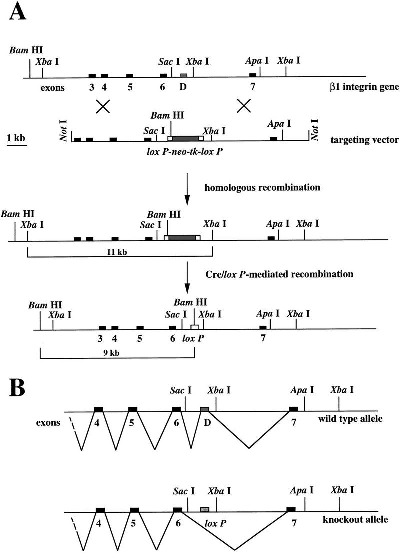
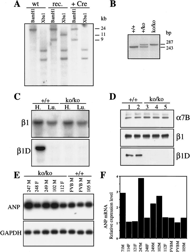
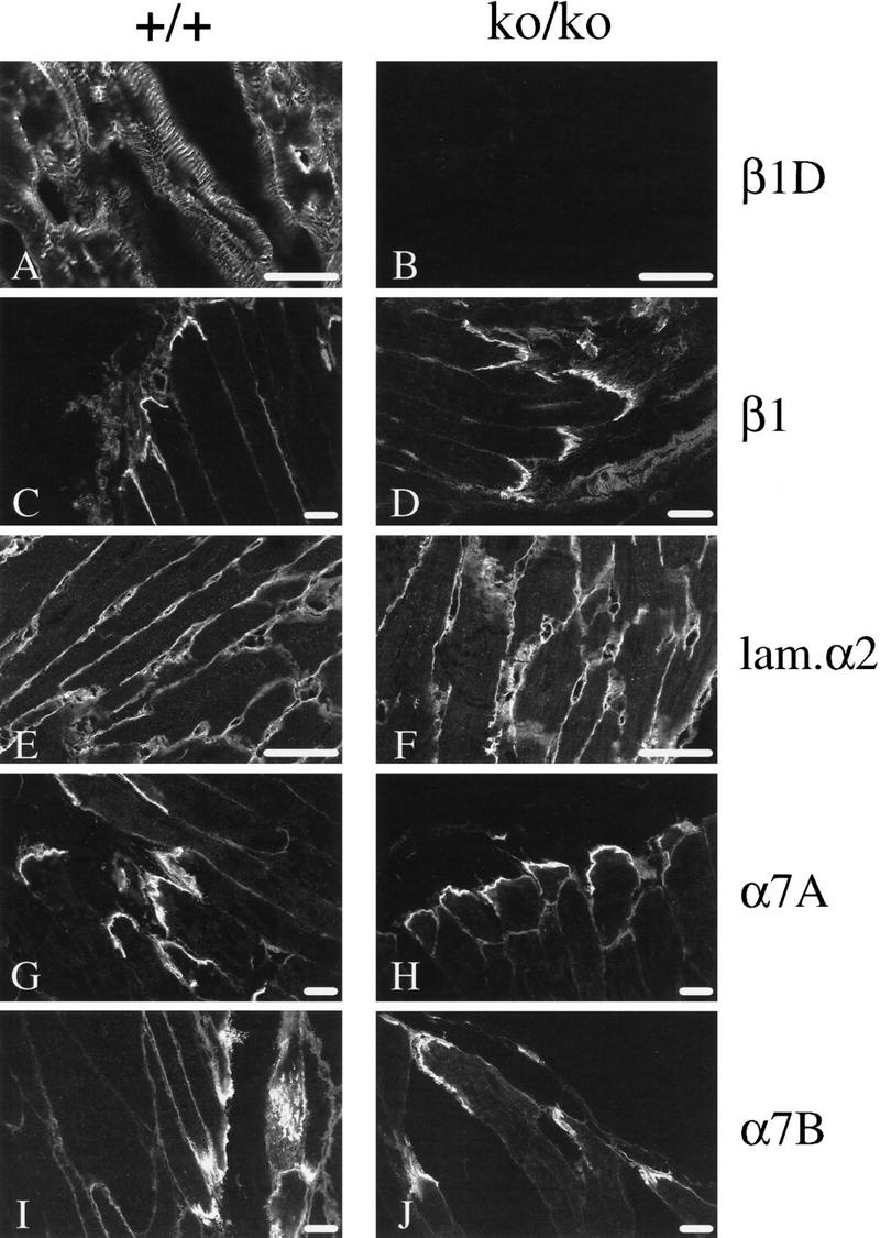
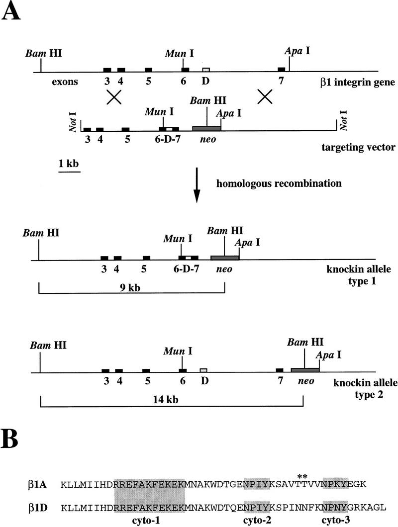
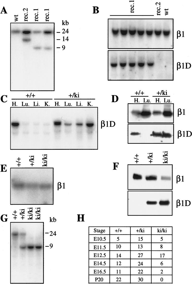
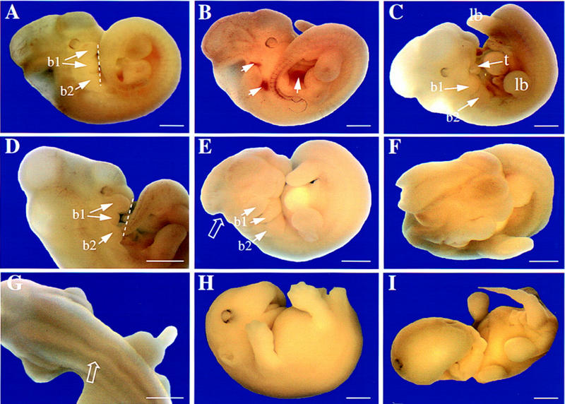
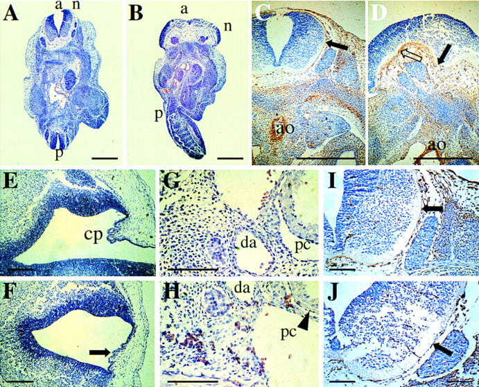
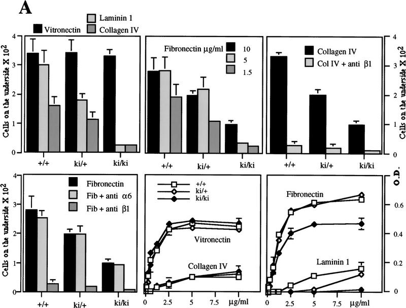
The beta1D integrin is a recently characterized isoform of the beta1 subunit that is specifically expressed in heart and skeletal muscle. In this study we have assessed the function of the beta1D integrin splice variant in mice by generating, for the first time, Cre-mediated exon-specific knockout and knockin strains for this splice variant. We show that removal of the exon for beta1D leads to a mildly disturbed heart phenotype, whereas replacement of beta1A by beta1D results in embryonic lethality with a plethora of developmental defects, in part caused by the abnormal migration of neuroepithelial cells. Our data demonstrate that the splice variants A and D are not functionally equivalent. We propose that beta1D is less efficient than beta1A in mediating the signaling that regulates cell motility and responses of the cells to mechanical stress.


Figures

Figure 1
Figure 1
Scheme for exon-specific targeting. (A) Restriction maps for the 3′ portion of the mouse β1 integrin locus, the targeting construct, the homologous recombinant, and the exon D-deleted mutant. In the targeting construct, exon D is replaced by a neo–tk minigene flanked by two loxP sites. After homologous recombination, targeted ES cells are transiently transfected with a Cre-encoding plasmid. After Cre/loxP-mediated recombination, exon D is replaced by a loxP site. Recombination is detected in Southern blot analysis as a new 9-kb BamHI fragment. The probe (331) is a cDNA consisting of exons 3, 4, 5, and 6. (B) In normal muscle cells, the exon D is inserted into the mature β1D mRNA, whereas in the knockout muscle cells, exon 6 and 7 are connected leading to the mature β1A mRNA.
Figure 2
Figure 2
(A) Identification of homologous and Cre/loxP recombination by Southern blot analysis of DNA from targeted ES cell lines. A 24-kb fragment is present in BamHI digested DNA from wild-type ES cells (wt), a 9-kb mutated fragment is also present in DNA from homologous-targeted ES cell before (rec) and after (+Cre) the Cre/loxP mediated recombination. In XbaI-digested DNA, an 11-kb fragment is present in the DNA from homologously targeted ES cells, whereas the fragment disappeared after transfection with the Cre-encoding plasmid, showing that the Cre–loxP recombination had been successful. (B) PCR analysis of tail genomic DNA from +/+, +/ko, and ko/ko β1D mice. Primers flank exon D; the lower band corresponds to exon D and its consensus splicing sites, the upper band corresponds to the loxP site. (C) Northern blot analysis of mRNA derived from heart and lungs of +/+ and β1D ko/ko mice. The blot is successively probed with an antisense oligonucleotide encoding exon D (β1D) and exon 7 (β1). (D) Immunoblot analysis of proteins extracted from the heart of +/+ and ko/ko mice. The blot is successively probed with anti-β1D, β1, and α7B antibodies. (E) Northern blot analysis of RNA extracted from ventricles of +/+ and ko/ko mice. The blot is probed successively with an ANP and GAPDH cDNA probes. (F) Quantification by PhosphorImager analysis of the relative expression level of ANP mRNA in heart ventricles of +/+ and ko/ko mice. (F) Female; (M) male.
Figure 3
Figure 3
(A–J) Immunostaining of skeletal muscles from β1D +/+ and ko/ko mice. Skeletal muscles of 1-year-old mice were frozen in OCT compound, and sections were prepared and subjected to immunofluorescence histochemistry with various antibodies. The first section of each pair is from +/+ skeletal muscles; the second is from ko/ko skeletal muscles. (A,B) anti-β1D; (C,D) anti-β1; (E,F) anti-α2 laminin subunit; (G,H) anti-α7A integrin; (I,J) anti-α7B integrin. Scale bars, 25 μm.
Figure 4
Figure 4
(A) Knockin targeting strategy. Restriction maps for the 3′ portion of the mouse β1 integrin locus, the targeting construct, and the type 1 and 2 homologous recombinants. Homologous recombination occurs between the 5′ and the 3′ end of the construct with the genomic DNA leading to the type 1 recombinant DNA or between the 3′ end and the connecting region between exon 7 and the neo gene with the genomic DNA leading to the type 2 recombinant DNA. After digestion with BamHI, the ES cell DNA is probed with 331, giving a wild-type 24-kb fragment and targeted 14- or 9-kb fragments. (B) The amino acid sequences of the β1A and β1D cytoplasmic domains differ after the KWDT sequence. The conserved cyto-1, cyto-2, and cyto-3, which are important for the localization of integrins in focal contacts, are shaded. Note that the two threonines (*) present in the β1A integrin are absent in the β1D integrin.
Figure 5
Figure 5
(A) Identification of type 1 and type 2 homologous recombination by Southern blot analysis of DNA from targeted ES cell lines. After digestion with BamHI, DNA is probed with 331. The 24-kb fragment is the wild-type allele (wt), the 9-kb and the 14-kb fragments are the type 1 (rec. 1) and type 2 (rec. 2) mutated alleles, respectively. (B) Northern blot analysis of RNA from total ES cells. The type 1-targeted ES cells (rec. 1) express β1D after homologous recombination. The blot was successively hybridized with an antisense oligonucleotide encoding for exon D (β1D) and exon 7 (β1). (C) Northern blot analysis of RNA from total tissues of heterozygous knockin mice. After knockin recombination, β1D is constitutively expressed in all the tissues analyzed, i.e., heart (H.), lungs (Lu.), liver (Li.), and kidneys (ki.). (D) Western blot analysis of proteins from total tissues of heterozygous knockin mice. Mice expressing β1D at the mRNA level (see Fig. 6C) express β1D at the protein level. (E) Northern blot analysis of total RNAs derived from knockin MEFs in culture. The wild-type +/+, heterozygous +/ki, and homozygous ki/ki β1D knockin MEFs express β1 mRNA at the same level. The blot was hybridized with probe 331, which recognizes all β1 isoforms. (F) Western blot analysis of proteins from a total lysate of MEFs. ki/ki MEFs express β1D. Note the higher expression level of β1 in the +/+ MEFs compared with the ki/ki MEFs. (G) Southern blot analysis of DNA derived from knockin embryos. In ki/ki embryos, only the type 1 mutated allele (9 kb) is present. The blot was hybridized with 331. (H) Genotypes of progeny from intercrosses between heterozygous β1D knockin parents.
Figure 6
Figure 6
Phenotype of β1D ki/ki embryos between 11.5 and 16.5 dpc. Gross morphology is shown at 11.5 (A–G) and 16.5 dpc (H,I). (A) Control +/ki embryo at 11.5 dpc. The neural tube is closed rostrally and caudally; brain cavities are evident as convolutions in the head; the tail bends to the right, with developing limbs symmetrically positioned on either side. The broken line joins ventral extremities of first and second branchial arches (b1, b2). In b1, the maxillary (top) and mandibular (bottom) components are already separating. (B) β1D ki/ki embryo at 11.5 dpc showing extravasation of RBCs (arrows). (C) β1D ki/ki embryo at 11.5 dpc showing that the maxillary component of the first branchial arch (b1) is missing, leaving the tongue (t) exposed. The limbs (lb) are excentrically positioned with respect to the tail. (D) β1D ki/ki embryo at 11.5 dpc showing abnormal development of the first branchial arch (b1) with the mandibular component missing. The broken line joins ventral extremities of b1 and b2. (E) β1D ki/ki embryo at 11.5 dpc showing lack of part of the hindbrain (open arrow). The body is also abnormally twisted with lower limbs excentrically placed relative to the tail and branchial arches b1 and b2 are underdeveloped. (F) Anterior view of embryo in E showing open neural tube. (G) Dorsal view of a β1D ki/ki embryo at 11.5 dpc showing a kinked neural tube (open arrow). (H) β1D ki/ki embryo at 16.5 dpc. The shortened branchial arch has resulted in an underdeveloped lower jaw, so that the upper palate is visible. The head is abnormally smooth. (F) β1D ki/ki embryo at 16.5 dpc. Both jaws and the lower part of the face are underdeveloped and misformed. One eye has been displaced to the top of the head, probably by edema (arrow). All limbs have retarded digit development. Scale bars, 1.2 mm.
Figure 7
Figure 7
Histological sections of control and β1D ki/ki embryos stained with hematoxilin and eosin. (A) Transverse section of control +/ki embryo (12.5 dpc) at the level of the hind limbs. The embryo is curved so that the anterior (a) and posterior (p) neural tubes (n) are visible and closed. (B) Transverse section of β1D ki/ki embryo (12.5 dpc) at the level of the hind limbs. The neural tube (n) is open and the neuroepithelium overexpanded. Twisting of the body results in an abnormal a–p axis. (C) FN staining of a control +/ki embryo (12.5 dpc) (transverse section through the anterior neural tube). Note the thin layer around the neural tube, the low level of staining in the mesenchyme ventral to the neural tube (solid arrow) and the strong staining around the aorta (ao). (D) FN staining of a β1D ki/ki embryo (12.5 dpc) (transverse section through the anterior neural tube that had failed to close). Note the lack of a thin layer of FN around the neural tube (open arrow), excessive FN in the mesenchyme underlying the neuroepithelium (solid arrow), but the normal amounts of FN around the aorta (a). (E) Sagittal section through the head of a control +/ki embryo at 14.5 dpc showing the choroid plexus (cp) in the roof of the fourth ventricle. (F) Sagittal section through the head of a β1D ki/ki embryo at 14.5 dpc at the position that should contain the choroid plexus (arrow). The head of this embryo is smooth, as in Fig. 6H. (G) Sagittal section through a control +/ki embryo showing RBCs confined within blood vessels [(da) dorsal aorta]. (H) Sagittal section through a β1D ki/ki embryo showing RBCs dispersed throughout the tissue and in the pericardial cavity (pc). (I) α5 integrin staining of a control +/ki embryo at 12.5 dpc. Transverse section adjacent in the anterior neural tube. Note staining of the same thin layer around the neural tube as in C. (J) α5 integrin staining of a β1D ki/ki embryo at 12.5 dpc. Transverse section through the anterior neural tube that had failed to close. The thin layer around the neural tube is stained as in controls in which the tube is closed at the same level. Scale bars, 1 mm in A–D, 150 μm E–J.
Figure 8
Figure 8
(A) Migratory (bars) and adhesive (lines) properties of +/+, +/ki, and ki/ki MEFs on collagen IV, laminin 1, vitronectin, and FN. By use of concentrations of FN that result in the same adhesion of +/+ (1.5 μg/ml) and ki/ki (5 μg/ml) MEFs (bottom right), migration is not restored to normal in the ki/ki MEFs. (Top right) Migration on collagen IV is completely blocked with an anti-β1 antibody. (Bottom left) Migration on FN is blocked completely by an anti-β1 antibody, whereas an anti-α6 antibody has no effect. (B) Immunoprecipitation analysis of various integrin subunits expressed at the surface of the +/+ and ki/ki MEFs. (Cont. Ab.) The secondary antibody used as a control.
Figure 8
Figure 8
(A) Migratory (bars) and adhesive (lines) properties of +/+, +/ki, and ki/ki MEFs on collagen IV, laminin 1, vitronectin, and FN. By use of concentrations of FN that result in the same adhesion of +/+ (1.5 μg/ml) and ki/ki (5 μg/ml) MEFs (bottom right), migration is not restored to normal in the ki/ki MEFs. (Top right) Migration on collagen IV is completely blocked with an anti-β1 antibody. (Bottom left) Migration on FN is blocked completely by an anti-β1 antibody, whereas an anti-α6 antibody has no effect. (B) Immunoprecipitation analysis of various integrin subunits expressed at the surface of the +/+ and ki/ki MEFs. (Cont. Ab.) The secondary antibody used as a control.

References

    1. Almeida EAC, Huovila APJ, Sutherland AE, Stephens LE, Calarco PG, Shaw LM, Mercurio AM, Sonnenberg A, Primakoff P, Myles DG, White JM. Mouse egg integrin α6β1 functions as a sperm receptor. Cell. 1995;81:1095–1104. - PubMed
    1. Altruda F, Cervella P, Tarone G, Botta C, Balzac F, Stefanuto G, Silengo L. A human integrin β1 subunit with a unique cytoplasmic domain generated by alternative mRNA processing. Gene. 1990;95:261–266. - PubMed
    1. Aumailley M, Mann K, von der Mark H, Timpl R. Cell attachment properties of collagen type IV and Arg-Gly-Asp dependent binding to its α2(VI) and α3(VI) chains. Exp Cell Res. 1989;181:463–474. - PubMed
    1. Baudoin C, Van der Flier A, Borradori L, Sonnenberg A. Genomic organization of the mouse β1 gene: Conservation of the β1D but not of the β1B and β1C integrin splice variants. Cell Adhes Commun. 1996;4:1–11. - PubMed
    1. Belkin AM, Zhidkova NI, Balzac F, Altruda F, Tomatis D, Maier A, Tarone G, Koteliansky VE, Burridge K. β1D integrin displaces the β1A isoform in striated muscles: Localization at junctional structures and signaling potential in nonmuscle cells. J Cell Biol. 1996;132:211–226. - PMC - PubMed

Publication types