. 1998 Apr;180(8):2050-6.
doi: 10.1128/JB.180.8.2050-2056.1998.

FtsZ dynamics during the division cycle of live Escherichia coli cells

Affiliations

FtsZ dynamics during the division cycle of live Escherichia coli cells

Q Sun et al. J Bacteriol. 1998 Apr.

Abstract

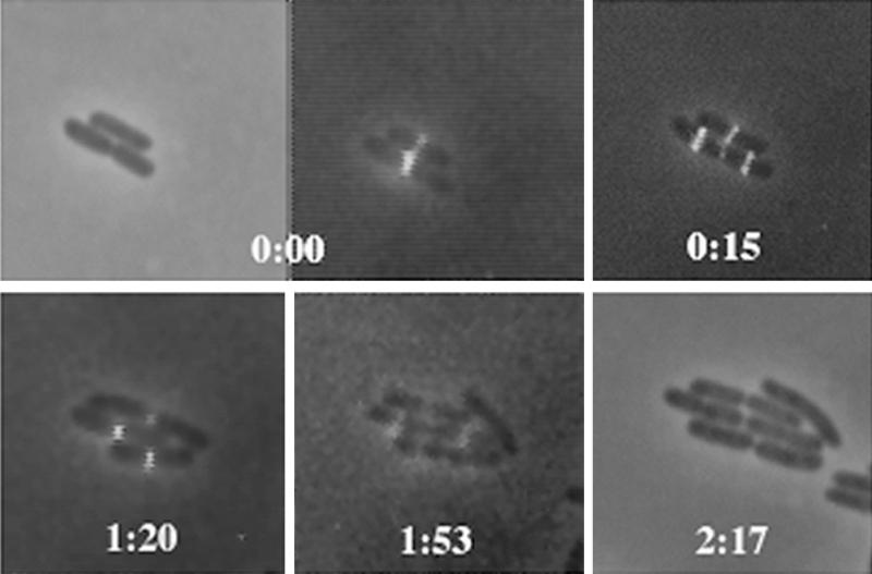
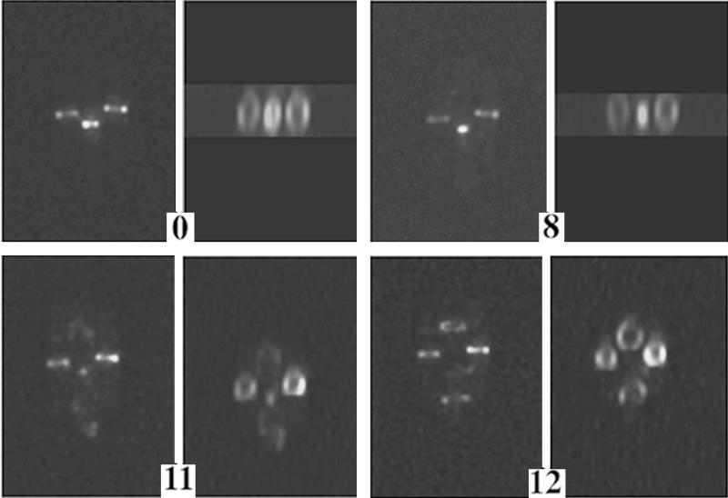
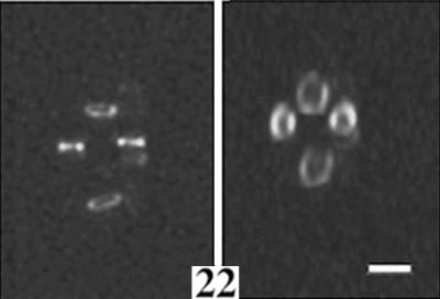
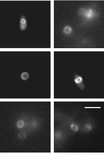
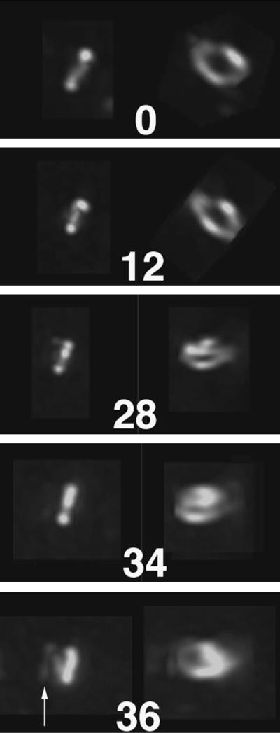
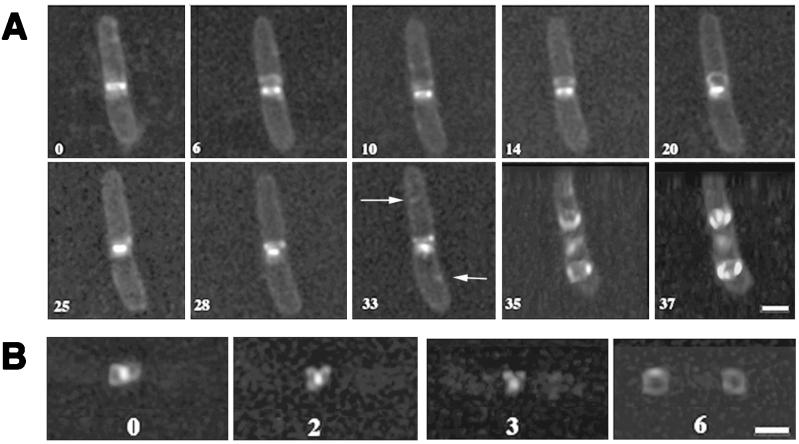
The dynamics and assembly of bacterial cell division protein FtsZ were monitored in individual, growing and dividing Escherichia coli cells in real time by microculture of a merodiploid strain expressing green fluorescent protein (GFP)-tagged FtsZ. Cells expressing FtsZ-GFP at levels less than or equivalent to that of wild-type FtsZ were able to grow and divide over multiple generations, with their FtsZ rings visualized by fluorescence. During the late stages of cytokinesis, which constituted the last one-fourth of the cell cycle, the lumen of the FtsZ ring disappeared as the whole structure condensed. At this time, loops of FtsZ-GFP polymers emanated outward from the condensing ring structure and other unstable fluorescent structures elsewhere in the cell were also observed. Assembly of FtsZ rings at new division sites occurred within 1 min, from what appeared to be single points. Interestingly, this nucleation often took place in the predivisional cell at the same time the central FtsZ ring was in its final contraction phase. This demonstrates directly that, at least when FtsZ-GFP is being expressed, new division sites have the capacity to become fully functional for FtsZ targeting and assembly before cell division of the mother cell is completed. The results suggest that the timing of FtsZ assembly may be normally controlled in part by cellular FtsZ concentration. The use of wide-field optical sectioning microscopy to obtain sharp fluorescence images of FtsZ structures is also discussed.


Figures

FIG. 1
FIG. 1
Growth and division of E. coli microcolonies expressing FtsZ-GFP and FtsZ, showing that FtsZ rings containing FtsZ-GFP function normally. Two consecutive cell division cycles are shown, using conventional fluorescence microscopy. The first and last panels are phase-contrast images; note that the last time point lacks a fluorescence image. The other panels were obtained by a digital overlay of phase-contrast and fluorescence images at the times shown. Times are shown in hours and minutes. Note the simultaneous formation of daughter FtsZ rings and contraction of the midcell ring at 0:15 in the bottom cell and at 1:53 in two daughter cells of the original bottom cell.
FIG. 2
FIG. 2
Three-dimensional image reconstruction of a shorter time course showing a fluorescence image of three adjacent growing cells, in vertical orientation, with the central cell undergoing complete septation and cell separation. Cells in the left-hand panels are viewed without rotation; the other panels show cells viewed with rotation to highlight the FtsZ annular structure as it condenses and duplicates. Times are shown in minutes; complete division of the central cell occurred between the 12- and 22-min time points. Note the formation of arcs in the 11-min panel that form nearly complete rings in the 12-min panel. Also note the fluorescence at the bottom pole of the dividing cell in the 11-min panel. Bar, 1.3 μm.
FIG. 2
FIG. 2
Three-dimensional image reconstruction of a shorter time course showing a fluorescence image of three adjacent growing cells, in vertical orientation, with the central cell undergoing complete septation and cell separation. Cells in the left-hand panels are viewed without rotation; the other panels show cells viewed with rotation to highlight the FtsZ annular structure as it condenses and duplicates. Times are shown in minutes; complete division of the central cell occurred between the 12- and 22-min time points. Note the formation of arcs in the 11-min panel that form nearly complete rings in the 12-min panel. Also note the fluorescence at the bottom pole of the dividing cell in the 11-min panel. Bar, 1.3 μm.
FIG. 3
FIG. 3
Visualization of FtsZ ring cross sections in immobilized, live cells. Shown are different cells, perpendicular to the plane of the coverslip, in which the FtsZ-GFP fluorescence displays a complete, uniform ring. Bar, 2 μm.
FIG. 4
FIG. 4
Formation of a spiral FtsZ-GFP polymer at midcell. Shown is a time course of a fluorescent FtsZ ring that ultimately fails to participate in septation and instead forms a clear spiral that emanates away from the midcell. The cell, expressing FtsZ-GFP, was grown in microculture and failed to divide over the several hours of growth. Times are shown in minutes. Images on the left for each time point are unrotated, whereas images on the right are rotated to reveal the cross-sectional structure. An arrow in the bottom panel highlights the developing spiral.
FIG. 5
FIG. 5
Depolymerization and reorganization of FtsZ during the last stages of septation, as shown by three-dimensional reconstruction. (A) A single growing cell during cytokinesis. To aid visualization of the cell outline, the membrane was stained with FM4-64 vital dye. Note the disappearance of the loop formed at 20 min by the 25-min time point, followed by reappearance at 28 min. Arrows point to putative nucleation sites for FtsZ assembly in daughter cells. Also note that at 35 min, the FtsZ structures were formed at the future daughter cell division sites while the old FtsZ structure was still located as a highly condensed form at midcell. In order to show FtsZ rings in the daughter cell more clearly, the cells at 35 and 37 min are shown at a tilted angle. (B) A more detailed picture of a single growing cell, showing dynamics of the FtsZ loops. The bright dot is the condensed FtsZ ring, and the rings at the 6-min time point are formed in the middles of daughter cells. The numbers throughout the figure represent the time course, in minutes, after the first image was acquired. Bars, 1.3 μm.

References

    1. Addinall S G, Bi E, Lutkenhaus J. FtsZ ring formation in fts mutants. J Bacteriol. 1996;178:3877–3884. - PMC - PubMed
    1. Addinall S G, Cao C, Lutkenhaus J. Temperature shift experiments with an ftsZ84(Ts) strain reveal rapid dynamics of FtsZ localization and indicate that the Z ring is required throughout septation and cannot reoccupy division sites once constriction has initiated. J Bacteriol. 1997;179:4277–4284. - PMC - PubMed
    1. Addinall S G, Lutkenhaus J. FtsZ-spirals and -arcs determine the shape of the invaginating septa in some mutants of Escherichia coli. Mol Microbiol. 1996;22:231–237. - PubMed
    1. Agard D A, Hiraoka Y, Shaw P, Sedat J W. Fluorescence microscopy in three dimensions. Methods Cell Biol. 1989;30:353–377. - PubMed
    1. Baumann P, Jackson S P. An archaebacterial homologue of the essential eubacterial cell division protein FtsZ. Proc Natl Acad Sci USA. 1996;93:6726–6730. - PMC - PubMed

Publication types

MeSH terms

LinkOut - more resources