. 1998 Apr;180(8):2102-9.
doi: 10.1128/JB.180.8.2102-2109.1998.

IS6110 transposition and evolutionary scenario of the direct repeat locus in a group of closely related Mycobacterium tuberculosis strains

Affiliations

IS6110 transposition and evolutionary scenario of the direct repeat locus in a group of closely related Mycobacterium tuberculosis strains

Z Fang et al. J Bacteriol. 1998 Apr.

Abstract

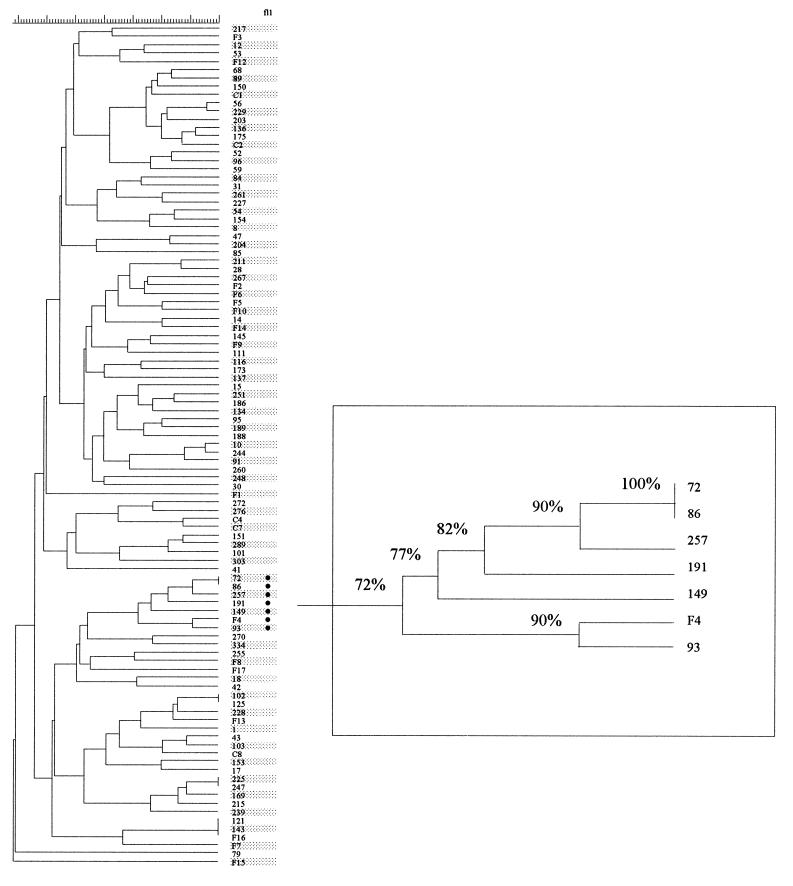
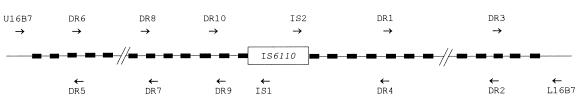
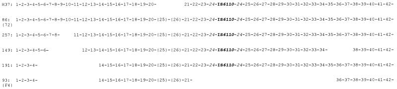
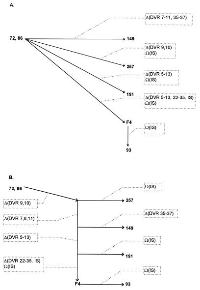
In recent years, various polymorphic loci and multicopy insertion elements have been discovered in the Mycobacterium tuberculosis genome, such as the direct repeat (DR) locus, the major polymorphic tandem repeats, the polymorphic GC-rich repetitive sequence, IS6110, and IS1081. These, especially IS6110 and the DR locus, have been widely used as genetic markers to differentiate M. tuberculosis isolates and will continue to be so used, due to the conserved nature of the genome of M. tuberculosis. However, little is known about the processes involved in generating these or of their relative rates of change. Without an understanding of the biological characteristics of these genetic markers, it is difficult to use them to their full extent for understanding the population genetics and epidemiology of M. tuberculosis. To address these points, we identified a cluster of 7 isolates in a collection of 101 clinical isolates and investigated them with various polymorphic genetic markers, which indicated that they were highly related to each other. This cluster provided a model system for the study of IS6110 transposition, evolution at the DR locus, and the effects of these on the determination of evolutionary relationships among M. tuberculosis strains. Our results suggest that IS6110 restriction fragment length polymorphism patterns are useful in grouping closely related isolates together; however, they can be misleading if used for making inferences about the evolutionary relationships between closely related isolates. DNA sequence analysis of the DR loci of these isolates revealed an evolutionary scenario, which, complemented with the information from IS6110, allowed a reconstruction of the evolutionary steps and relationships among these closely related isolates. Loss of the IS6110 copy in the DR locus was noted, and the mechanisms of this loss are discussed.


Figures

FIG. 1
FIG. 1
Dendrogram of IS6110 fingerprints of 101 isolates of M. tuberculosis, constructed with GelCompar and based on Dice coefficients and UPGMA clustering. In column fl1, the symbol (•) indicates isolates with fl1::IS6110. The graph in the box is an enlarged branch of the isolates carrying fl1::IS6110 and shows the Dice similarity coefficients between the isolates.
FIG. 2
FIG. 2
PCR amplification of the fl1::IS6110 allele. (A) PCR products obtained with primers FL1a and FL1b from an fl1+ strain (lane 1) and an fl1::IS6110 strain (lane 2). Lane 3, 1-kb ladder. (B) IS6110 (open box) and fl1 (shaded boxes), with the locations of the primers used and directions of their extension.
FIG. 3
FIG. 3
IS6110, DR, and FL1 RFLP patterns of fl1::IS6110 isolates. Isolates F4, 93, 72, 86, 149, 257, and 191 were digested with PvuII and, following blotting, were probed sequentially with IS6110 (), DR (⇐), and FL1 (←) probes. Lane M, internal size standards, with DNA sizes marked in kilobase pairs.
FIG. 4
FIG. 4
Autoradiogram of PvuII-digested DNA from M. tuberculosis isolates hybridized with the PGRS probe (A) and the IS1547 probe (B). Lanes 1 through 9, isolates H37Ra, 72, 86, 93, 149, 191, 257, F4, and Mt14323, respectively. Sizes are indicated on the right, in kilobases.
FIG. 5
FIG. 5
Schematic illustration of the PCR primers used in this study. The filled boxes represent the DR sequences, between which are spacer DNA sequences. The locations and directions of extension of the primers are indicated.
FIG. 6
FIG. 6
Schematic representation of the structures of the DR regions of M. tuberculosis isolates. A number and the hyphen following it represent one DVR. The DVRs are numbered from the 5′ end to the 3′ end of the DNA sequence. The DR structure of M. tuberculosis H37Rv (H37) (EMBL accession no. Z81331) is shown for reference. The numbers in parentheses, i.e., (25) and (26), represent DVR spacers identical to the spacers of DVR25 and DVR26 in M. bovis BCG (EMBL accession no. X57835) (19). IS6110 is inserted into DVR24, which is therefore shown on each side of the insertion.
FIG. 7
FIG. 7
Deduced evolutionary scenarios for the seven closely related isolates, inferred from their DR locus polymorphisms and their IS6110 distributions. Solid lines with arrows represent routes; dotted lines and dotted boxes indicate changes of DRs and/or IS6110. Route A is based on the assumption that the structures of the DR loci of these isolates are the results of independent events, while route B assumes that they result from sequential events. Δ and Ω, deletion and insertion, respectively.

References

    1. Achtman M. A surfeit of YATMs? J Clin Microbiol. 1996;34:1870. . (Letter.) - PMC - PubMed
    1. Altschul S F. Amino acid substitution matrices from an information theoretic perspective. J Mol Biol. 1991;219:555–565. - PMC - PubMed
    1. Altschul S F, Gish W, Miller W, Myers E W, Lipman D J. Basic local alignment search tool. J Mol Biol. 1990;215:403–410. - PubMed
    1. Ayala F J, Barrio E, Kwiatowski J. Molecular clock or erratic evolution? A tale of two genes. Proc Natl Acad Sci USA. 1996;93:11729–11734. - PMC - PubMed
    1. Bloom B R. Tuberculosis. Back to a frightening future. Nature. 1992;358:538–539. - PubMed

Publication types

LinkOut - more resources