Transformation of Rickettsia prowazekii to rifampin resistance
- PMID: 9555894
- PMCID: PMC107138
- DOI: 10.1128/JB.180.8.2118-2124.1998
Transformation of Rickettsia prowazekii to rifampin resistance
Erratum in
- J Bacteriol 1998 Aug;180(15):4015
Abstract
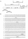
Rickettsia prowazekii, the causative agent of epidemic typhus, is an obligate intracellular parasitic bacterium that grows directly within the cytoplasm of the eucaryotic host cell. The absence of techniques for genetic manipulation hampers the study of this organism's unique biology and pathogenic mechanisms. To establish the feasibility of genetic manipulation in this organism, we identified a specific mutation in the rickettsial rpoB gene that confers resistance to rifampin and used it to demonstrate allelic exchange in R. prowazekii. Comparison of the rpoB sequences from the rifampin-sensitive (Rifs) Madrid E strain and a rifampin-resistant (Rifr) mutant identified a single point mutation that results in an arginine-to-lysine change at position 546 of the R. prowazekii RNA polymerase beta subunit. A plasmid containing this mutation and two additional silent mutations created in codons flanking the Lys-546 codon was introduced into the Rifs Madrid E strain of R. prowazekii by electroporation, and in the presence of rifampin, resistant rickettsiae were selected. Transformation, via homologous recombination, was demonstrated by DNA sequencing of PCR products containing the three mutations in the Rifr region of rickettsial rpoB. This is the first successful demonstration of genetic transformation of Rickettsia prowazekii and represents the initial step in the establishment of a genetic system in this obligate intracellular pathogen.
Figures


References
-
- Aliabadi Z, Winkler H H, Wood D O. Isolation and characterization of the Rickettsia prowazekii gene encoding the flavoprotein subunit of succinate dehydrogenase. Gene. 1993;133:135–140. - PubMed
-
- Andersson S G E, Eriksson A-S, Näslund A K, Andersen M S, Kurland C G. The Rickettsia prowazekii genome: a random sequence analysis. Microb Comp Genomics. 1997;1:293–315. - PubMed
-
- Ausubel F, Brent R, Kingston R E, Moore D D, Seidman J G, Smith J A, Struhl K, editors. Current protocols in molecular biology. 1, 2, and 3. New York, N.Y: John Wiley & Sons, Inc.; 1997.
-
- Balayeva N M, Frolova O M, Genig V A, Nikolskaya V N. Some biological properties of antibiotic resistant mutants of Rickettsia prowazekii strain E induced by nitroso-guanidine. In: Kazár J, editor. Rickettsiae and rickettsial diseases. Bratislava, Slovakia: Publishing House of the Slovak Academy of Sciences; 1985. pp. 85–91.
Publication types
MeSH terms
Substances
Associated data
- Actions
Grants and funding
LinkOut - more resources
Full Text Sources
Other Literature Sources
Medical

