. 1998 Apr;180(8):2232-6.
doi: 10.1128/JB.180.8.2232-2236.1998.

A novel DNA polymerase family found in Archaea

Affiliations

A novel DNA polymerase family found in Archaea

Y Ishino et al. J Bacteriol. 1998 Apr.

Abstract

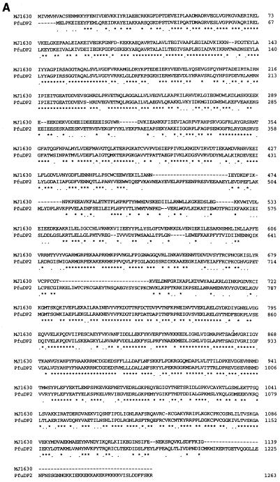
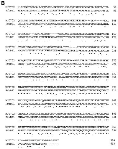
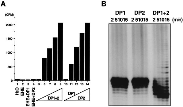
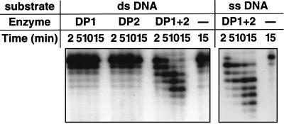
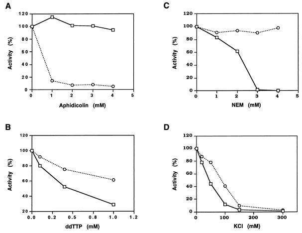
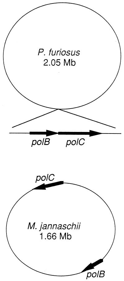
One of the most puzzling results from the complete genome sequence of the methanogenic archaeon Methanococcus jannaschii was that the organism may have only one DNA polymerase gene. This is because no other DNA polymerase-like open reading frames (ORFs) were found besides one ORF having the typical alpha-like DNA polymerase (family B). Recently, we identified the genes of DNA polymerase II (the second DNA polymerase) from the hyperthermophilic archaeon Pyrococcus furiosus, which has also at least one alpha-like DNA polymerase (T. Uemori, Y. Sato, I. Kato, H. Doi, and Y. Ishino, Genes Cells 2:499-512, 1997). The genes in M. jannaschii encoding the proteins that are homologous to the DNA polymerase II of P. furiosus have been located and cloned. The gene products of M. jannaschii expressed in Escherichia coli had both DNA polymerizing and 3'-->5' exonuclease activities. We propose here a novel DNA polymerase family which is entirely different from other hitherto-described DNA polymerases.


Figures

FIG. 1
FIG. 1
Alignment of the two components of DNA Pol II from P. furiosus and M. jannaschii. (A and B) DP2 and DP1, respectively. CLUSTAL W (13) was used for sequence alignment of the two polymerases. Identical and similar amino acid residues at the same positions are indicated by asterisks and dots, respectively, in both alignments.
FIG. 1
FIG. 1
Alignment of the two components of DNA Pol II from P. furiosus and M. jannaschii. (A and B) DP2 and DP1, respectively. CLUSTAL W (13) was used for sequence alignment of the two polymerases. Identical and similar amino acid residues at the same positions are indicated by asterisks and dots, respectively, in both alignments.
FIG. 2
FIG. 2
Detection of the DNA polymerizing activity from the MJ0702 and MJ1630 proteins. The reaction conditions are described in the text. (A) The acid-insoluble radioactivity bound to DE 81 paper detected by a scintillation counter. Protein samples are indicated under the bar graph as follows: EHE, HE from BL21(DE3)/pET21a; DP1, HE from BL21(DE3)/pMJDP1; DP2, HE from BL21(DE3)/pMJDP2. Lanes 1 and 5, H2O; lane 2, 14 μg; lanes 3 and 4, 7.0 μg each; lane 6, 1.75 μg each; lane 7, 3.5 μg each; lane 8, 5.25 μg each; lane 9, 7.0 μg each; lanes 10 to 14, DP1 (7.0 μg) and various amounts of DP2 (lane 10, 0 μg; lane 11, 1.75 μg; lane 12, 3.5 μg; lane 13, 5.25 μg; and lane 14, 7.0 μg). (B) Chain elongation ability detected by a primer extension reaction with a single-stranded DNA annealed with a 32P-labeled d40-mer (described in the text) as a template primer. Equal amounts were sampled at 2, 5, 10, and 15 min from the reaction mixture and loaded onto an 8% polyacrylamide gel containing 8 M urea. The electrophoretic profile was visualized by autoradiography.
FIG. 3
FIG. 3
Detection of the 3′→5′ exonuclease activity from the MJ0702 and MJ1630 proteins. For the 3′→5′ exonuclease assay, equal aliquots of the reaction mixture described in the text were removed after 2, 5, 10, and 15 min and were added to a stop solution. Products were analyzed by polyacrylamide gel electrophoresis in the presence of 8 M urea as described for Fig. 2B. ds, double stranded; ss, single stranded.
FIG. 4
FIG. 4
Inhibition of the DNA polymerizing activity of M. jannaschii Pol II (DP1-DP2) by aphidicolin, ddTTP, N-ethylmaleimide (NEM), and salt. The standard assay mixture containing [methyl-3H]TTP for DNA polymerizing activity as described in the text was processed in the presence of the indicated amounts of inhibitors. Acid-insoluble radioactivities were counted with a scintillation counter. The open squares and circles indicate M. jannaschii Pol II and P. furiosus Pol I, respectively.
FIG. 5
FIG. 5
Localization of the two genes corresponding to the new DNA polymerase in the genomes of P. furiosus and M. jannaschii. The locations of the genes in M. jannaschii follow the published map. The location of the P. furiosus operon is indeterminate, due to the absence of a complete genomic sequence.

Similar articles

Cited by

References

    1. Brush G S, Kelly T J. Mechanisms for replicating DNA. In: DePamphilis M L, editor. DNA replication in eukaryotic cells. Cold Spring Harbor, N.Y: Cold Spring Harbor Laboratory Press; 1996. pp. 1–43.
    1. Bult C J, White O, Olsen G J, Zhou L, Fleischmann R D, Sutton G G, Blake J A, FitzGerald L M, Clayton R A, Gocayne J D, Kerlavage A R, Dougherty B A, Tomb J-F, Adams M D, Reich C I, Overbeek R, Kirkness E F, Weinstock K G, Merrick J M, Glodek A, Scott J L, Geoghagen N S M, Weidman J F, Fuhrmann J L, Presley E A, Nguyen D, Utterback T R, Kelley J M, Peterson J D, Sadow P W, Hanna M C, Cotton M D, Hurst M A, Roberts K M, Kaine B P, Borodovsky M, Klenk H-P, Fraser C M, Smith H O, Woese C R, Venter J C. Complete genome sequence of the methanogenic archaeon, Methanococcus jannaschii. Science. 1996;273:1058–1073. - PubMed
    1. Edgell D R, Doolittle W F. Archaebacterial genomics: the complete genome sequence of Methanococcus jannaschii. Bioessays. 1996;19:1–4.
    1. Edgell D R, Doolittle W F. Archaea and the origin(s) of DNA replication proteins. Cell. 1997;89:995–998. - PubMed
    1. Edgell D R, Klenk H-P, Doolittle W F. Gene duplications in evolution of archaeal family B DNA polymerases. J Bacteriol. 1997;179:2632–2640. - PMC - PubMed

Substances

LinkOut - more resources