. 1998 May;72(5):3524-33.
doi: 10.1128/JVI.72.5.3524-3533.1998.

Activation of caspases in pig kidney cells infected with wild-type and CrmA/SPI-2 mutants of cowpox and rabbitpox viruses

Affiliations

Activation of caspases in pig kidney cells infected with wild-type and CrmA/SPI-2 mutants of cowpox and rabbitpox viruses

J Macen et al. J Virol. 1998 May.

Abstract

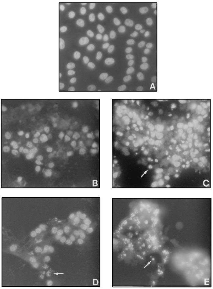
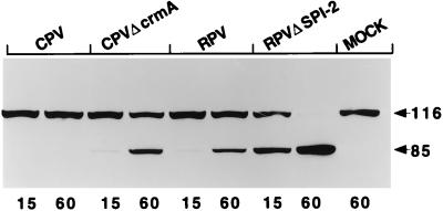
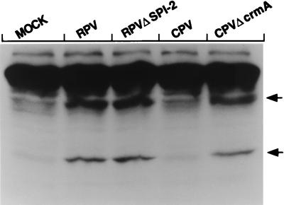
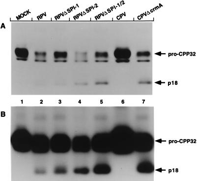
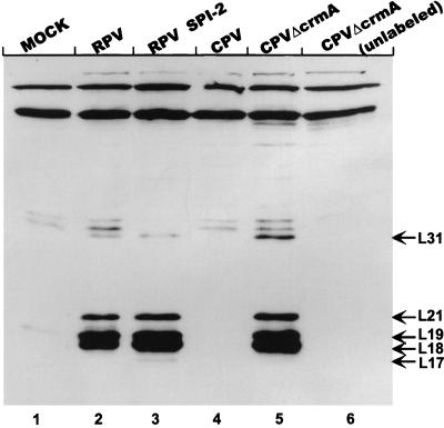
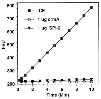
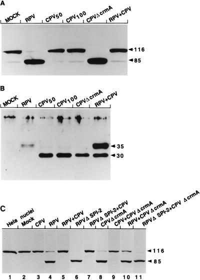
The cowpox virus (CPV) CrmA and the equivalent rabbitpox virus (RPV) SPI-2 proteins have anti-inflammatory and antiapoptosis activity by virtue of their ability to inhibit caspases, including the interleukin-1beta-converting enzyme (ICE; caspase-1). Infection of LLC-PK1 pig kidney cells with a CPV CrmA mutant, but not with wild-type (wt) CPV, results in the induction of many of the morphological features of apoptosis (C. A. Ray and D. J. Pickup, Virology 217:384-391, 1996). In our study, LLC-PK1 cells infected with CPV delta crmA, but not those infected with wt CPV, showed induction of poly(ADP-ribose) polymerase (PARP)- and lamin A-cleaving activities and processing of the CPP32 (caspase-3) precursor to a mature 18-kDa form. Surprisingly, infection of LLC-PK1 cells with either wt RPV (despite the presence of the SPI-2 protein) or RPV delta SPI-2 resulted in cleavage activity against PARP and lamin A and the appearance of the mature subunit of CPP32/caspase-3. The biotinylated specific peptide inhibitor Ac-Tyr-Val-Lys(biotinyl)-Asp-2,6-dimethylbenzoyloxymethylketone [AcYV(bio)KD-aomk] labeled active caspase subunits of 18, 19, and 21 kDa in extracts from LLC-PK1 cells infected with CPV delta crmA, wt RPV, or RPV delta SPI-2 but not wt CPV. Mixed infection of LLC-PK1 cells with wt RPV and wt CPV gave no PARP-cleaving activity, and all PARP cleavage mediated by SPI-2 and CrmA mutants of RPV and CPV, respectively, could be eliminated by coinfection with wt CPV. These results suggest that the RPV SPI-2 and CPV CrmA proteins are not functionally equivalent and that CrmA, but not SPI-2 protein, can completely prevent apoptosis in LLC-PK1 cells under these conditions.


Figures

FIG. 1
FIG. 1
DAPI staining of CPV- or RPV-infected LLC-PK1 cells. Cells were mock infected (A) or infected with wt CPV (B), CPVΔcrmA (C), wt RPV (D), or RPVΔSPI-2 (E) and then stained with DAPI at 16 h postinfection as described in Materials and Methods. The arrows indicate examples of apoptotic nuclei.
FIG. 2
FIG. 2
PARP-cleaving activity in infected LLC-PK1 cells. Extracts prepared from LLC-PK1 cells at 14 h postinfection were mixed with purified HeLa cell nuclei, and the mixtures were incubated for either 15 or 60 min (indicated by the numbers below the lanes) at 37°C. Proteins from the reaction were separated on SDS–10% polyacrylamide gels, and PARP was detected by immunoblotting with the anti-PARP monoclonal antibody C-2-10. The arrows indicate the intact 116-kDa molecule and the 85-kDa cleavage product. MOCK, mock-infected cell extract.
FIG. 3
FIG. 3
Human lamin A-cleaving activity in extracts from infected LLC-PK1 cells. Extracts prepared from LLC-PK1 cells at 14 h postinfection were mixed with 35S-labeled human lamin A, produced by in vitro transcription-translation, and incubated for 90 min at 37°C. The proteins were resolved by SDS–10% PAGE and visualized by autoradiography. The arrows indicate the two fragments resulting from lamin A cleavage. MOCK, mock-infected cell extract.
FIG. 4
FIG. 4
Extracts from wt CPV-infected cells do not inhibit PARP-cleaving activity. Extracts prepared from infected LLC-PK1 cells at 14 h postinfection were mixed with purified HeLa cell nuclei, and the mixtures were incubated for 60 min at 37°C. Proteins from the reaction were separated by SDS–10% PAGE, and PARP was detected by immunoblotting with the monoclonal antibody C-2-10. In the odd-numbered lanes are extracts prepared from singly infected cells, mixed and preincubated at 37°C for 30 min before the nuclei were added. In the even-numbered lanes are the nuclear extracts that were mixed and added directly to the nuclei with no preincubation. The arrows indicate the intact 116-kDa molecule and the 85-kDa cleavage product. MOCK, mock-infected cell extract.
FIG. 5
FIG. 5
Processing of caspase-3 (CPP32) in extracts from infected LLC-PK1 cells. LLC-PK1 cells were either mock infected (MOCK) or infected with one of the following: RPV, RPV with a mutation in either the SPI-1 gene (a serpin with approximately 50% identity to SPI-2), the SPI-2 gene, or both SPI-1 and SPI-2; wild-type CPV; or the CPV CrmA mutant. Extracts prepared at 14 h postinfection were resolved on an SDS–15% polyacrylamide gel, and caspase-3 was detected by immunoblotting with a polyclonal rabbit antiserum directed against the p17 subunit of human caspase-3. (A) Blot with short exposure period; (B) longer exposure of the same blot shown in panel A. The arrows indicate the positions of the unprocessed proenzyme from porcine cells (pro-CPP32) and the cleaved subunit which runs at 18 kDa on SDS gels (p18).
FIG. 6
FIG. 6
Labelling of activated caspases in infected-cell extracts with Ac-YV(bio)KD-aomk. Lanes 1 to 5, extracts prepared from mock-infected (MOCK) or infected LLC-PK1 cells at 14 h postinfection, labelled with the biotinylated tetrapeptide inhibitor Ac-YV(bio)KD-aomk as described in Materials and Methods; lane 6, extract from CPVΔcrmA-infected cells incubated in the absence of the tetrapeptide. Proteins from the reactions were resolved on SDS–16% polyacrylamide gels. Labelled caspases were detected with streptavidin-HRP followed by ECL. Bands corresponding to labelled caspases were designated L followed by numbers indicating their approximate molecular masses (kilodaltons) and are indicated with arrows. This figure was prepared by using Adobe Photoshop 3.0 software for the Macintosh computer and an Epson GT-9500 scanner.
FIG. 7
FIG. 7
Expression of CrmA and SPI-2 in infected LLC-PK1 cells. LLC-PK1 cells were infected with either wt RPV or wt CPV at an MOI of 5 PFU/cell (RPV) or 25 PFU/cell (CPV). At the times indicated (in hours) above the lanes, extracts were prepared from the infected cells and an equal amount of protein from each was loaded onto an SDS–12% polyacrylamide gel. SPI-2 and CrmA were detected with the SPI-2- and CrmA-specific monoclonal antibody 2B12-4B1. The arrow indicates the position of the 38-kDa SPI-2/CrmA protein. In the RPV-infected cells, a lower-molecular-mass protein is also occasionally visualized at late times and may represent a minor cross-reacting protein or a further degradation product of SPI-2.
FIG. 8
FIG. 8
Inhibition of ICE by both purified CrmA and purified SPI-2. His-tagged CrmA and SPI-2 proteins were purified as described in Materials and Methods, preincubated with purified human recombinant ICE for 5 min, and then incubated with a fluorogenic ICE substrate (Ac-YVAD-AMC). Fluorescence readings were taken at the indicated times and plotted as arbitrary fluorescence signal units (FSU).
FIG. 9
FIG. 9
PARP-cleaving activity in LLC-PK1 cells coinfected with RPV and CPV derivatives. Extracts were prepared from cells that had been either mock infected (MOCK), singly infected with RPV (MOI = 10 [A and B] or 15 [C]), CPV (MOI = 50 [CPV50] [A and B], 100 [CPV100] [A and B], or 15 [C]), CPVΔcrmA (MOI = 15 [A and B] or 5 [C]), or RPVΔSPI-2 (MOI = 5 [C]), or dually infected with two of the following: CPV (MOI = 15), RPV (MOI = 15), CPVΔcrmA (MOI = 5), and RPVΔSPI-2 (MOI = 5). All samples were harvested at 14 h postinfection. (A) Extracts were assayed for PARP-cleaving activity, using purified HeLa cell nuclei and immunoblotting with the anti-PARP monoclonal antibody. The arrows indicate the intact 116-kDa molecule and the 85-kDa cleavage fragment. (B) Extracts were separated on an SDS–12% polyacrylamide gel and immunoblotted with a polyclonal rabbit antiserum specific for the orthopoxvirus chemokine-binding protein. The CPV version of this protein migrates at approximately 30 kDa, while the RPV homolog migrates at 35 kDa, as indicated by the arrows. (C) PARP immunoblot of HeLa cell nuclei that either were untreated (lane 1) or had been incubated with extracts from mock-infected (lane 2) or infected (lanes 3 to 11) cells. The viruses used to generate the various extracts are indicated above the lanes.

Similar articles

Cited by

References

    1. Alcami A, Smith G L. Cytokine receptors encoded by poxviruses: a lesson in cytokine biology. Immunol Today. 1995;16:474–478. - PubMed
    1. Alcami A, Smith G L. Soluble interferon-gamma receptors encoded by poxviruses. Comp Immunol Microbiol Infect Dis. 1996;19:305–317. - PubMed
    1. Ali A N, Turner P C, Brooks M A, Moyer R W. The SPI-1 gene of rabbitpox virus determines host range and is required for hemorrhagic pock formation. Virology. 1994;202:306–314. - PubMed
    1. Alnemri E S, Livingston D J, Nicholson D W, Salvesen G, Thornberry N A, Wong W W, Yuan J. Human ICE/CED-3 protease nomenclature. Cell. 1996;87:171. - PubMed
    1. Boldin M P, Goncharov T M, Goltsev Y V, Wallach D. Involvement of MACH, a novel MORT1/FADD-interacting protease, in Fas/APO-1- and TNF receptor-induced cell death. Cell. 1996;85:803–815. - PubMed

Publication types

MeSH terms