. 1998 May;72(5):3554-9.
doi: 10.1128/JVI.72.5.3554-3559.1998.

Elongation of the cytoplasmic tail interferes with the fusion activity of influenza virus hemagglutinin

Affiliations

Elongation of the cytoplasmic tail interferes with the fusion activity of influenza virus hemagglutinin

M Ohuchi et al. J Virol. 1998 May.

Abstract

The hemagglutinin (HA) of fowl plague virus was lengthened and shortened by site-specific mutagenesis at the cytoplasmic tail, and the effects of these modifications on HA functions were analyzed after expression from a simian virus 40 vector. Elongation of the tail by the addition of one to six histidine (His) residues did not interfere with intracellular transport, glycosylation, proteolytic cleavage, acylation, cell surface expression, and hemadsorption. However, the ability to induce syncytia at a low pH decreased dramatically depending on the number of His residues added. Partial fusion (hemifusion), assayed by fluorescence transfer from octadecylrhodamine-labeled erythrocyte membranes, was also reduced, but even with the mutant carrying six His residues, significant transfer was observed. However, when the formation of fusion pores was examined with hydrophilic fluorescent calcein, transfer from erythrocytes to HA-expressing cells was not observed with the mutant carrying six histidine residues. The addition of different amino acids to the cytoplasmic tail of HA caused an inhibitory effect similar to that caused by the addition of His. On the other hand, a mutant lacking the cytoplasmic tail was still able to fuse at a reduced level. These results demonstrate that elongation of the cytoplasmic tail interferes with the formation and enlargement of fusion pores. Thus, the length of the cytoplasmic tail plays a critical role in the fusion process.


Figures
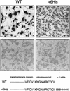
FIG. 1
FIG. 1
Hemadsorption (bottom panels) and cell fusion (top panels) of CV-1 cells expressing WT HA and +6His HA. Cells were infected with recombinant SV40-HA stocks and incubated for 2 days. HA was expressed in the presence of 10 mM ammonium chloride for protection against acid degradation during transport (20). Before hemadsorption, HA-expressing cells were treated with VCNA to remove sialic acid interfering with the binding of erythrocytes (21). Cell fusion was induced by acid treatment (pH 5.0). Amino acid sequences of the cytoplasmic tail of the WT and the +6His mutant are also shown.
FIG. 2
FIG. 2
Specific fusion activity of the WT and +His mutants. HA expression and fusion tests were done as described in the legend to Fig. 1. The fusion index is the total number of nuclei in syncytia per field divided by the number of hemadsorption-positive cells per field before fusion induction.
FIG. 3
FIG. 3
Posttranslational processing of WT HA and mutant HA. (A) Proteolytic cleavage of HA. Expressed HA was labeled with [35S]methionine, recovered by immunoprecipitation, and then analyzed by PAGE. (B) Endo H digestion of HA. Labeled HA was recovered as described above and digested with endo H. (C) Acylation of HA. Expressed HA was labeled with [3H]palmitic acid for 16 h and analyzed by PAGE without 2-mercaptoethanol.
FIG. 4
FIG. 4
Fluorescence transfer from R18-labeled erythrocyte membranes to HA-expressing cells. After adsorption of R18-labeled erythrocytes, HA-expressing cells were treated at pH 5.0 for 5 min, and then the transfer of fluorescence was immediately examined under a fluorescence microscope. Photographs were taken 10 min after fusion induction. HK, Hong Kong type HA used as a control.
FIG. 5
FIG. 5
Fluorescence transfer from calcein-filled erythrocytes to HA-expressing cells. Hemadsorption and fusion tests were done as described in the legend to Fig. 1. Photographs were taken 10 min after fusion induction.

Similar articles

Cited by

References

    1. Bullough P A, Hughson F M, Skehel J J, Wiley D C. Structure of influenza haemagglutinin at the pH of membrane fusion. Nature. 1994;371:37–43. - PubMed
    1. Crawford S, Goff S P. A deletion mutation in the 5′ part of the pol gene of Moloney murine leukemia virus blocks proteolytic processing of the Gag and Pol polyproteins. J Virol. 1985;53:899–907. - PMC - PubMed
    1. Fass D, Harrison S C, Kim P S. Retrovirus envelope domain at 1.7 angstrom resolution. Nat Struct Biol. 1996;3:465–469. - PubMed
    1. Garten W, Will C, Buckard K, Kuroda K, Ortmann D, Munk K, Scholtissek C, Schnittler H, Drenckhahn D, Klenk H-D. Structure and assembly of hemagglutinin mutants of fowl plague virus with impaired surface transport. J Virol. 1992;66:1495–1505. - PMC - PubMed
    1. Green N, Shinnick T M, Witte O, Ponticelli A, Sutcliffe J G, Lerner R A. Sequence-specific antibodies show that maturation of Moloney leukemia virus envelope polyprotein involves removal of a COOH-terminal peptide. Proc Natl Acad Sci USA. 1981;78:6023–6027. - PMC - PubMed

Publication types

MeSH terms

LinkOut - more resources