. 1998 May;72(5):3571-7.
doi: 10.1128/JVI.72.5.3571-3577.1998.

Coding sequences enhance internal initiation of translation by hepatitis A virus RNA in vitro

Affiliations

Coding sequences enhance internal initiation of translation by hepatitis A virus RNA in vitro

J Graff et al. J Virol. 1998 May.

Abstract

Hepatitis A virus (HAV), unlike other picornaviruses, has a slow-growth phenotype in permissive cell lines and in general does not induce host cell cytopathology. Although there are no published reports of productive infection of HeLa cells by HAV, HAV RNA appears to be readily translated in HeLa cells when transcribed by T7 RNA polymerase provided by a recombinant vaccinia virus. The 5' noncoding region of HAV was fused to poliovirus (PV) coding sequences to determine the effect on translation efficiency in HeLa cell extracts in vitro. Conditions were optimized for utilization of the HAV internal ribosome entry segment (IRES). Transcripts from chimeric constructs fused precisely at the initiation codon were translated very poorly. However, chimeric RNAs which included 114 or more nucleotides from the HAV capsid coding sequences downstream of the initiation codon were translated much more efficiently than those lacking these sequences, making HAV-directed translation efficiency similar to that directed by the PV IRES. Sixty-six nucleotides were insufficient to confer increased translation efficiency. The most 5'-terminal HAV 138 nucleotides, previously determined to be upstream of the IRES, had an inhibitory effect on translation efficiency. Constructs lacking these terminal sequences, or those in which the PV 5'-terminal sequences replaced those from HAV, translated three- to fourfold better than those with the intact HAV 5'-terminal end.


Figures
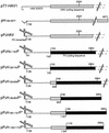
FIG. 1
FIG. 1
Schematic representation of the chimeric HAV/PV constructs used to examine the efficiency of HAV IRES-driven translation. All constructs contain the promoter for T7 RNA polymerase. The striped lines represent the HAV 5′NCR starting at the indicated position, nt 1, 91, or 139; solid lines show the PV 5′-terminal cloverleaf structure from nt 1 to 109. Striped boxes represent HAV coding sequences until nt 806, 854, 1191, 1194, or 4977 or until the 3′ end of the HAV genome if not indicated; shaded boxes represent PV coding sequences starting at nt 743, 746, or 1172 fused in frame to HAV sequences. Nucleotide numbering of the HAV genome corresponds to the wild-type HAV strain HM175 (9). The 5′NCR and the coding region are not in actual proportion to each other.
FIG. 2
FIG. 2
Western immunoblot analysis of HAV and PV proteins expressed in HeLa cells. Proteins generated from plasmids pPsHAV (A, lanes 1 to 3), pT7-HAV1 (A, lanes 4 to 6), pPsPV1 (B, lanes 1 to 3), and pT7PV1 (B, lanes 4 to 6) were analyzed by SDS-PAGE 24 h posttransfection in the presence of recombinant vaccinia virus vTF7-3, carrying the gene for T7 RNA polymerase. The proteins were transferred to a nitrocellulose membrane and detected by HAV VP1 or PV VP2 antiserum. Each plasmid DNA was used at 3, 6, and 9 μg for transfection. Extract from mock-transfected HeLa cells as negative control (lane 7) and prestained molecular weight markers (M, lane 8) were analyzed on the same gel. Sizes of marker proteins are indicated on the right in kilodaltons.
FIG. 3
FIG. 3
Translation in vitro of truncated HAV RNAs with different 5′-terminal sequences. Decreasing RNA amounts (1, 0.5, and 0.25 μg) of transcripts derived from pT7-HAV1, pH139-4977, and pPsHAV, linearized with BstZ17I (nt 2024), were used to program HeLa cell translation extracts in the presence of a mixture of [35S]methionine and [35S]cysteine. The translation products were analyzed by SDS-PAGE and subjected to autoradiography. The predicted size of each product was calculated to be ∼47 kDa as indicated. PV-encoded polypeptides obtained from PV-infected HeLa cells labeled with [35S]methionine served as protein marker (M, lane 1) and are identified on the left. Lane 2 shows background translation products from the HeLa cell extracts with no added RNA.
FIG. 4
FIG. 4
Translation of truncated HAV and HAV/PV chimeric RNAs in HeLa cell extracts. Translation products from 500 ng of the indicated RNA per 12.5-μl reaction mixture were resolved by SDS-PAGE and subjected to autoradiography. Lane 1 (M) represents PV-encoded polypeptides obtained from PV-infected HeLa cells labeled with [35S]methionine, identified on the left. PV RNA (100 ng), isolated from purified PV virions (lane 2), was used as an internal translation control. The predicted molecular masses of the translation products derived from transcripts HAV1* and PsHAV*, linearized at nt 2024 (47 kDa), and from transcripts PsH91-740P*, PsH139-740P*, and PsH1194/1172P*, linearized at nt 2954 (81 kDa), are indicated.
FIG. 5
FIG. 5
Translation of HAV/PV RNAs with extended HAV capsid coding sequences downstream of the HAV 5′NCR. HeLa cell extract was programmed with 500 and 250 ng of transcript derived from pPsHAV, truncated at nt 2024, or pPsH139-740P, pPsH139-806P, pPsH139-854P*, and pPsH139-1191P*, truncated at nt 2954, in the presence of [35S]methionine and [35S]cysteine. Translation products were resolved by SDS-PAGE and subjected to autoradiography. Lane 1 (M) represents PV-encoded polypeptides obtained from PV-infected HeLa cells labeled with [35S]methionine. The PV proteins used as marker are identified on the left. Lane 12 represents the negative control without addition of RNA, and lane 13 represents the internal translation control prepared by using PV RNA isolated from purified PV virions.

Similar articles

Cited by

References

    1. Alexander L, Lu H-H, Wimmer E. Poliovirus containing picornavirus type 1 and/or type 2 internal ribosomal entry site elements: genetic hybrids and the expression of a foreign gene. Proc Natl Acad Sci USA. 1994;91:1406–1410. - PMC - PubMed
    1. Anderson D A, Ross B C. Morphogenesis of hepatitis A virus: isolation and characterization of subviral particles. J Virol. 1990;64:5284–5289. - PMC - PubMed
    1. Andino R, Silvera D, Suggett S D, Achacoso P L, Miller C J, Baltimore D, Feinberg M B. Engineering poliovirus as a vaccine vector for the expression of diverse antigens. Science. 1994;265:1448–1451. - PubMed
    1. Barton D J, Flanegan J B. Coupled translation and replication of poliovirus RNA in vitro: synthesis of functional 3D polymerase and infectious virus. J Virol. 1993;67:822–831. - PMC - PubMed
    1. Blyn L B, Towner J S, Semler B L, Ehrenfeld E. Requirement of poly(rC) binding protein 2 for translation of poliovirus RNA. J Virol. 1997;71:6243–6246. - PMC - PubMed

Publication types

LinkOut - more resources