. 1998 May;72(5):3595-601.
doi: 10.1128/JVI.72.5.3595-3601.1998.

Monoclonal antibodies to distinct sites on herpes simplex virus (HSV) glycoprotein D block HSV binding to HVEM

Affiliations

Monoclonal antibodies to distinct sites on herpes simplex virus (HSV) glycoprotein D block HSV binding to HVEM

A V Nicola et al. J Virol. 1998 May.

Abstract

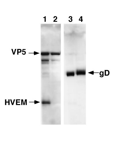
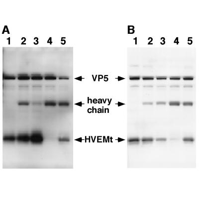
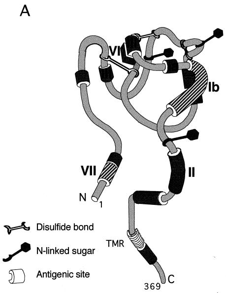
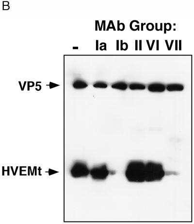
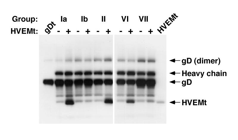
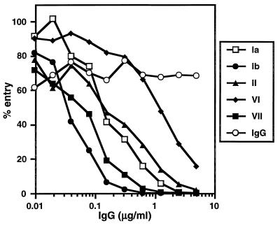
HVEM (for herpesvirus entry mediator) is a member of the tumor necrosis factor receptor superfamily and mediates entry of many strains of herpes simplex virus (HSV) into normally nonpermissive Chinese hamster ovary (CHO) cells. We used sucrose density centrifugation to demonstrate that purified HSV-1 KOS virions bind directly to a soluble, truncated form of HVEM (HVEMt) in the absence of any other cell-associated components. Therefore, HVEM mediates HSV entry by serving as a receptor for the virus. We previously showed that soluble, truncated forms of HSV glycoprotein D (gDt) bind to HVEMt in vitro. Here we show that antibodies specific for gD, but not the other entry glycoproteins gB, gC, or the gH/gL complex, completely block HSV binding to HVEM. Thus, virion gD is the principal mediator of HSV binding to HVEM. To map sites on virion gD which are necessary for its interaction with HVEM, we preincubated virions with gD-specific monoclonal antibodies (MAbs). MAbs that recognize antigenic sites Ib and VII of gD were the only MAbs which blocked the HSV-HVEM interaction. MAbs from these two groups failed to coprecipitate HVEMt in the presence of soluble gDt, whereas the other anti-gD MAbs coprecipitated HVEMt and gDt. Previous mapping data indicated that site VII includes amino acids 11 to 19 and site Ib includes 222 to 252. The current experiments indicate that these sites contain residues important for HSV binding to HVEM. Group Ib and VII MAbs also blocked HSV entry into HVEM-expressing CHO cells. These results suggest that the mechanism of neutralization by these MAbs is via interference with the interaction between gD in the virus and HVEM on the cell. Group Ia and II MAbs failed to block HSV binding to HVEM yet still neutralized HVEM-mediated entry, suggesting that these MAbs block entry at a step other than HVEM binding.


Figures

FIG. 1
FIG. 1
Cosedimentation of HSV-1 with soluble HVEMt. HVEMt (150 μg) and purified HSV-1 KOS (lanes 1 and 3) or rid1 (lanes 2 and 4) virions (107 PFU) were incubated for 2 h at 4°C and then passed through a 10%-30%-60% sucrose step gradient. The virus-containing band was collected from the 30%-60% sucrose interface and analyzed by SDS-PAGE (12% polyacrylamide) followed by Western blotting with anti-VP5 and anti-HVEM polyclonal Abs (lanes 1 and 2) or anti-gD polyclonal Ab (lanes 3 and 4). Secondary Abs were then added, and enhanced chemiluminescence was used to visualize the bands.
FIG. 2
FIG. 2
Inhibition of HSV-1 binding to HVEMt by Abs specific for HSV envelope glycoproteins. Gels in panels A and B represent two separate experiments carried out in the same way. Purified HSV-1 KOS virions (107 PFU) were preincubated for 1 h at 37°C with the following Abs or left untreated (−). The Abs specific for gB were SS10 (0.5 μl of ascites), DL16 (5 μg of IgG), DL21 (5 μg of IgG), and R69 (0.5 μl of sera) (lane 2). The Abs specific for gC were MP1 (0.5 μl of ascites), MP5 (5 μg of IgG), 1C8 (5 μg of IgG), and R46 (0.5 μl of sera) (lane 3). The Abs specific for gD were 1D3 (0.5 μl of ascites), DL2 (5 μg of IgG), DL11 (5 μg of IgG), and R7 (0.5 μl of sera) (lane 4). The Abs specific for gH/gL were LP11 (0.5 μl of ascites), 53S (5 μg of IgG), H6 (5 μg of IgG), and R137 (0.5 μl of sera) (lane 5). Samples were then subjected to sedimentation with HVEMt as described in the legend to Fig. 1. Samples were analyzed by SDS-PAGE (12% polyacrylamide) followed by Western blotting with anti-VP5 and anti-HVEM polyclonal Abs. Secondary Abs were then added, and enhanced chemiluminescence was used to visualize the bands.
FIG. 3
FIG. 3
Model of gD antigenic structure and inhibition of HSV-1 binding to HVEMt by a panel of anti-gD MAbs. (A) Hypothetical folded model of gD based on epitope mapping studies (37) and solution of the disulfide bond arrangement (32). Antigenic sites relevant to this study are indicated by Roman numerals. The approximate amino acid location of each site is as follows. Site Ia includes residues 216 to 234, site Ib includes residues 222 to 252, site II includes residues 272 to 279, site III includes residues 21 to 226, site VI includes residues 21 to 226, and site VII includes residues 11 to 19. Sites with hatch marks (Ib and VII) are important for HSV binding to HVEM. (B) Purified HSV-1 KOS virions were preincubated with 50 μg of the MAb IgGs HD1 (group Ia), DL11 (group Ib), DL6 (group II), DL2 (group VI), and 1D3 (group VII) or left untreated (lane 1) for 1 h at 37°C. Samples were then subjected to sedimentation with HVEMt as described in the legend to Fig. 1. Samples were analyzed by SDS-PAGE (12% polyacrylamide) followed by Western blotting with anti-VP5 and anti-HVEM polyclonal Abs. Secondary Abs were then added, and enhanced chemiluminescence was used to visualize the bands. TMR, transmembrane region.
FIG. 3
FIG. 3
Model of gD antigenic structure and inhibition of HSV-1 binding to HVEMt by a panel of anti-gD MAbs. (A) Hypothetical folded model of gD based on epitope mapping studies (37) and solution of the disulfide bond arrangement (32). Antigenic sites relevant to this study are indicated by Roman numerals. The approximate amino acid location of each site is as follows. Site Ia includes residues 216 to 234, site Ib includes residues 222 to 252, site II includes residues 272 to 279, site III includes residues 21 to 226, site VI includes residues 21 to 226, and site VII includes residues 11 to 19. Sites with hatch marks (Ib and VII) are important for HSV binding to HVEM. (B) Purified HSV-1 KOS virions were preincubated with 50 μg of the MAb IgGs HD1 (group Ia), DL11 (group Ib), DL6 (group II), DL2 (group VI), and 1D3 (group VII) or left untreated (lane 1) for 1 h at 37°C. Samples were then subjected to sedimentation with HVEMt as described in the legend to Fig. 1. Samples were analyzed by SDS-PAGE (12% polyacrylamide) followed by Western blotting with anti-VP5 and anti-HVEM polyclonal Abs. Secondary Abs were then added, and enhanced chemiluminescence was used to visualize the bands. TMR, transmembrane region.
FIG. 4
FIG. 4
Coimmunoprecipitation of HVEMt by a panel of anti-gD MAbs. Fifty-microliter reaction mixtures of 3 μg of gDt per ml in the presence (+) or absence (−) of 16 μg of HVEMt per ml were incubated for 1 h on ice. MAb ascites (0.1 μl) of HD1 (group Ia), DL11 (group Ib), DL6 (group II), DL2 (group VI), or 1D3 (group VII) were added for 1 h, followed by 50 μl of protein A-agarose (50 mg/ml) for 1 h. Bound material was collected by centrifugation at 13,000 × g for 3 min. Pellets were washed and then analyzed by SDS-PAGE (12% polyacrylamide). Western blots were probed with anti-gD and anti-HVEM polyclonal Abs. Secondary Abs were then added, and enhanced chemiluminescence was used to visualize the bands. Lane 1, gDt alone as a standard; lane 12, HVEMt alone as a standard.
FIG. 5
FIG. 5
Neutralization of HSV-1 entry by anti-gD MAb IgG. Purified HSV-1 KOS(tk12) was incubated with twofold dilutions of the MAb IgGs HD1 (group Ia), DL11 (group Ib), DL6 (group II), DL2 (group VI), and 1D3 (group VII) or nonimmune mouse IgG for 1 h at 37°C. Confluent CHO(250-2) cell monolayers on 96-well plates were infected with virus-Ab mixtures (4 × 10⁴ PFU per well) for 1 h at 4°C and then shifted to 37°C for 7 h to allow for virus entry. Nonidet P-40 (0.1%) cell lysates were prepared, and then substrate was added, and β-galactosidase activity (milli-optical density units per minute) was read at 560 nm. One hundred percent entry corresponds to β-galactosidase activity in the absence of IgG. Each point represents the average of duplicate wells. Shown are the results of one representative experiment. The experiment was repeated three times with similar results.

Similar articles

Cited by

References

    1. Baker S J, Reddy E P. Transducers of life and death: TNF receptor superfamily and associated proteins. Oncogene. 1996;12:1–9. - PubMed
    1. Bosch D L, Geerligs H J, Weijer W J, Feijlbrief M, Welling G W, Welling-Wester S. Structural properties and reactivity of N-terminal synthetic peptides of herpes simplex virus type 1 glycoprotein D by using antipeptide antibodies and group VII monoclonal antibodies. J Virol. 1987;61:3607–3611. - PMC - PubMed
    1. Brunetti C R, Burke R L, Kornfeld S, Gregory W, Masiarz F R, Dingwell K S, Johnson D C. Herpes simplex virus glycoprotein D acquires mannose 6-phosphate residues and binds to mannose 6-phosphate receptors. J Biol Chem. 1994;269:17067–17074. - PubMed
    1. Buckmaster E A, Gompels U, Minson A. Characterisation and physical mapping of an HSV-1 glycoprotein of approximately 115 × 10(3) molecular weight. Virology. 1984;139:408–413. - PubMed
    1. Campadelli-Fiume G, Arsenakis M, Farabegoli F, Roizman B. Entry of herpes simplex virus 1 in BJ cells that constitutively express viral glycoprotein D is by endocytosis and results in degradation of the virus. J Virol. 1988;62:159–167. - PMC - PubMed

Publication types

MeSH terms