. 1998 May;72(5):3658-65.
doi: 10.1128/JVI.72.5.3658-3665.1998.

The roles of Pol and Env in the assembly pathway of human foamy virus

Affiliations

The roles of Pol and Env in the assembly pathway of human foamy virus

D N Baldwin et al. J Virol. 1998 May.

Abstract

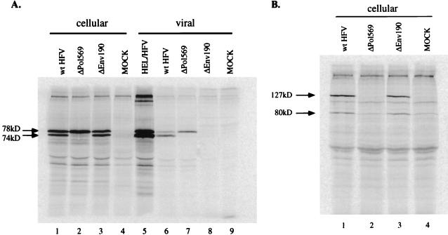
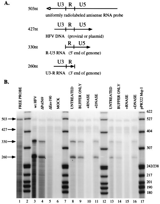
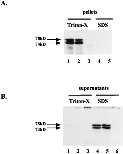
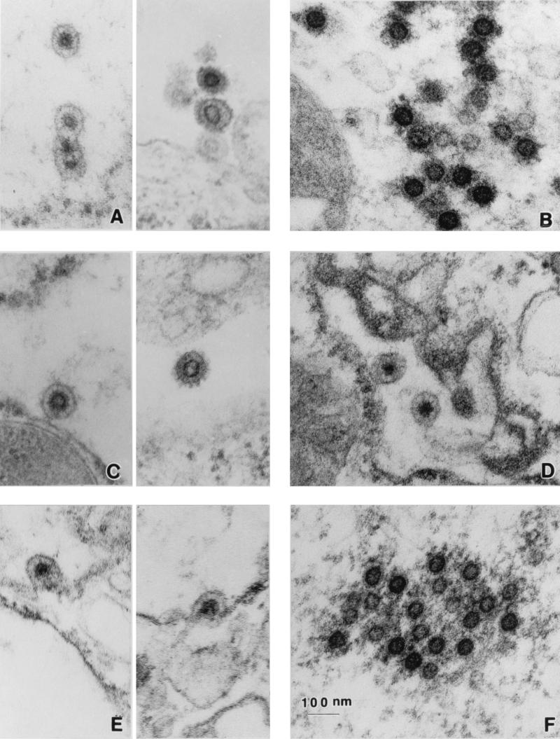
Human foamy virus (HFV) is the prototype of the Spumavirus genus of retroviruses. These viruses have a genomic organization close to that of other complex retroviruses but have similarities to hepadnaviruses such as human hepatitis B virus (HBV). Both HFV and HBV express their Pol protein independently of their structural proteins. Retroviruses and hepadnaviruses differ in their requirements for particle assembly and genome packaging. Assembly of retroviral particles containing RNA genomes requires only the Gag structural protein. The Pol protein is not required for capsid assembly, and the Env surface glycoprotein is not required for release of virions from the cell. In contrast, assembly of extracellular HBV particles containing DNA requires core structural protein and polymerase (P protein) for assembly of nucleocapsids and requires surface glycoproteins for release from the cell. We investigated the requirements for synthesis of extracellular HFV particles by constructing mutants with either the pol or env gene deleted. We found that the Pol protein is dispensable for production of extracellular particles containing viral nucleic acid. In the absence of Env, intracellular particles are synthesized but few or no extracellular particles could be detected. Thus, foamy virus assembly is distinct from that of other reverse transcriptase-encoding mammalian viruses.


Figures

FIG. 1
FIG. 1
RIPA of cellular and viral Gag and Pol proteins. Assay conditions and antibodies are described in Materials and Methods. (A) RIPA with anti-Gag antiserum. Lanes 1 to 4 contain cellular Gag proteins from transfected cells. Lanes: 1, wild-type (wt) HFV; 2, ΔPol569; 3, ΔEnv190; 4, mock-transfected FAB cells. Lane 5 contains virus from the supernatant of labelled, acutely infected HEL cells. Lanes 6 to 9 contain extracellular viral Gag proteins from the supernatants of the transfections. Lanes: 6, wild-type HFV; 7, ΔPol569; 8, ΔEnv190; 9, mock-transfected cells. Lanes 6 and 7 were used to normalize the number of wild-type HFV and ΔPol569 particles for the packaging experiments. (B) RIPA with anti-Pol antiserum. Cellular lysates for this figure were identical to those used with the anti-Gag antiserum. Lanes: 1, wild-type HFV; 2, ΔPol569; 3, ΔEnv190; 4, mock-transfected cells.
FIG. 2
FIG. 2
RPA of wild-type and mutant HFV nucleic acids. (A) The pSGC11 RNA probe is depicted, along with the predicted fragments protected by viral nucleic acids. The open arrow depicts the antisense probe, and the solid arrow represents the direction of viral transcription. (B) Results of RPA. Lanes 2, 7, 12, and 17 contain pBR322 MspI-digested, radiolabelled marker DNA. Other lanes: 1, 1% of total undigested free probe; 3, untreated wild-type (wt) HFV; 4, untreated ΔPol569; 5, untreated ΔEnv190; 6, mock-transfected cell supernatant; 8, untreated wild-type HFV; 9, mock-digested wild-type HFV; 10, RNase-treated wild-type HFV; 11, DNase-treated wild-type HFV; 13, untreated ΔPol569; 14, mock-digested ΔPol569; 15, RNase-treated ΔPol569; 16, DNase-treated ΔPol569. Lanes 3 to 6 were used for quantitation.
FIG. 3
FIG. 3
Comparison of intracellular particle formation by wild-type and ΔEnv190 by using Western blot analysis. Cell lysates were prepared as discussed in Materials and Methods. (A) Pellets. Lanes: 1 to 3, Triton X-lysed cells; 4 and 5, SDS-lysed cells. Lanes 1 and 4 contain wild-type HFV; lanes 2 and 5 contain ΔEnv190; lane 3 contains mock-transfected cells. (B) Supernatants. Lanes: 1 to 3, Triton X-lysed cells; 4 to 6, SDS-lysed cells. Lanes 1 and 4 contain wild-type HPV; lanes 2 and 5 contain ΔEnv190; lanes 3 and 6 contain mock-transfected cells.
FIG. 4
FIG. 4
TEM analysis of wild-type and mutant HFV particles. (A and B) Representative examples of wild-type HFV-transfected cells. (C) ΔPol569-transfected cells. (D to F) ΔEnv190-transfected cells. For wild-type (A) and ΔPol569 (C) enveloped particles, intracellular virions are shown on the left and extracellular virions are shown on the right. Cytoplasmic capsids are shown only for wild-type (B) and ΔEnv190 (F) capsids. Descriptions for fixing and staining of transfected FAB cells can be found in Materials and Methods.

Comment in

References

    1. Achong B G, Mansell W A, Epstein M A, Clifford P. An unusual virus in cultures from a human nasopharyngeal carcinoma. J Natl Cancer Inst. 1971;46:299–307. - PubMed
    1. Aronoff R, Hajjar A M, Linial M L. Avian retroviral RNA encapsidation: reexamination of functional 5′ RNA sequences and the role of nucleocapsid Cys-His motifs. J Virol. 1993;67:178–188. - PMC - PubMed
    1. Bartenschlager R, Junker-Niepmann M, Schaller H. The P gene product of hepatitis B virus is required as a structural component for genomic RNA encapsidation. J Virol. 1990;64:5324–5332. - PMC - PubMed
    1. Bartenschlager R, Schaller H. Hepadnavirus assembly is initiated by polymerase binding to the encapsidation signal in the viral RNA genome. EMBO J. 1992;11:3413–3420. - PMC - PubMed
    1. Birnbaum F, Nassal M. Hepatitis B virus nucleocapsid assembly: primary structure requirements in the core protein. J Virol. 1990;64:3319–3330. - PMC - PubMed

Publication types

LinkOut - more resources