

Substitution of ras for the herpesvirus saimiri STP oncogene in lymphocyte transformation
- PMID: 9557651
- PMCID: PMC109591
- DOI: 10.1128/JVI.72.5.3698-3704.1998
Substitution of ras for the herpesvirus saimiri STP oncogene in lymphocyte transformation
Abstract
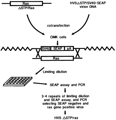
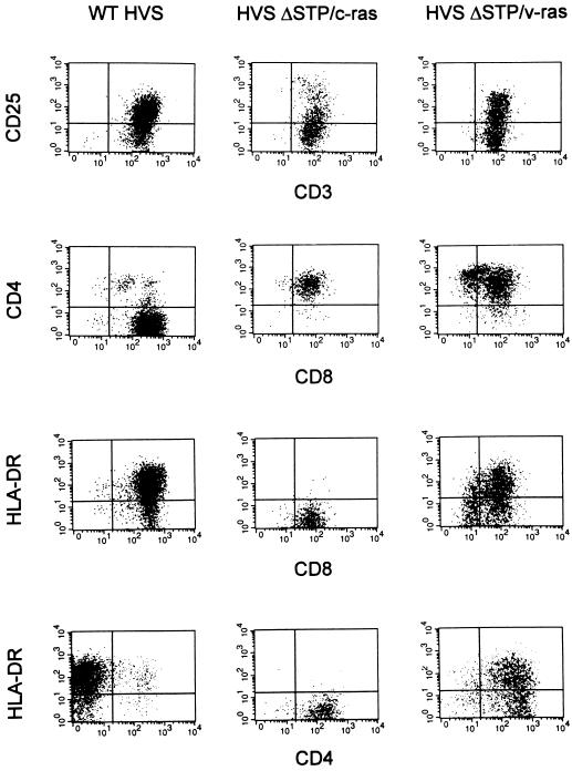
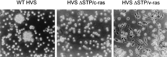
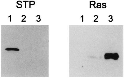
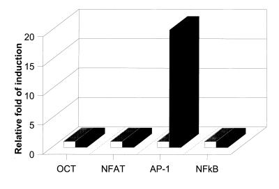
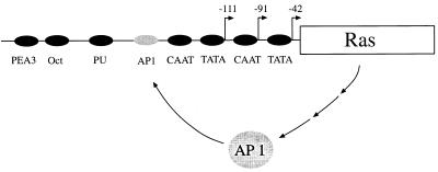
STP-C488 (STP of herpesvirus saimiri [HVS] group C strain 488 [C488]) is the only virus-encoded protein found to associate with cellular ras and activate ras signal transduction pathways. To investigate an important role for ras signal transduction in STP-dependent growth transformation, we constructed recombinant strains of HVS C488 in which the STP-C488 oncogene was replaced with cellular normal ras (c-ras) or viral oncogenic ras (v-ras). Recombinant HVS deltaSTP/v-ras immortalized primary common marmoset T lymphocytes to interleukin-2-independent growth as efficiently as wild-type HVS C488 (wt HVS), while recombinant HVS deltaSTP/c-ras did so with low efficiency. Whereas wt HVS immortalized CD4- CD8+ single-positive T lymphocytes, HVS deltaSTP/c-ras- and HVS deltaSTP/v-ras-immortalized cells were principally CD4+ CD8+ double-positive T lymphocytes. In addition, HVS deltaSTP/v-ras-immortalized T cells showed a high level of ras expression and exhibited an adherent macrophage-like morphology. These phenotypes were likely caused by the drastic activation of AP-1 transcriptional factor activity. Finally, HVS deltaSTP/v-ras and HVS deltaSTP/c-ras each induced lymphoma in one of two common marmosets, although onset of disease was more rapid with the v-ras virus. These results demonstrate that ras can substitute for the STP oncogene of HVS C488 to allow immortalized growth of primary lymphoid cells and that an activated form of ras does so more efficiently than the normal cellular form of ras.
Figures
Similar articles
-
Herpesvirus saimiri.Philos Trans R Soc Lond B Biol Sci. 2001 Apr 29;356(1408):545-67. doi: 10.1098/rstb.2000.0780. Philos Trans R Soc Lond B Biol Sci. 2001. PMID: 11313011 Free PMC article. Review.
-
STP and Tip are essential for herpesvirus saimiri oncogenicity.J Virol. 1998 Feb;72(2):1308-13. doi: 10.1128/JVI.72.2.1308-1313.1998. J Virol. 1998. PMID: 9445031 Free PMC article.
-
Role of cellular tumor necrosis factor receptor-associated factors in NF-kappaB activation and lymphocyte transformation by herpesvirus Saimiri STP.J Virol. 1999 May;73(5):3913-9. doi: 10.1128/JVI.73.5.3913-3919.1999. J Virol. 1999. PMID: 10196286 Free PMC article.
-
A role for herpesvirus saimiri orf14 in transformation and persistent infection.J Virol. 1998 Aug;72(8):6770-6. doi: 10.1128/JVI.72.8.6770-6776.1998. J Virol. 1998. PMID: 9658125 Free PMC article.
-
Herpesvirus saimiri as a model for gammaherpesvirus oncogenesis.Semin Cancer Biol. 1999 Jun;9(3):231-9. doi: 10.1006/scbi.1998.0115. Semin Cancer Biol. 1999. PMID: 10343074 Review.
Cited by
-
Herpesvirus ateles Tio can replace herpesvirus saimiri StpC and Tip oncoproteins in growth transformation of monkey and human T cells.J Virol. 2004 Sep;78(18):9814-9. doi: 10.1128/JVI.78.18.9814-9819.2004. J Virol. 2004. PMID: 15331715 Free PMC article.
-
Downregulation of p56(lck) tyrosine kinase activity in T cells of squirrel monkeys (Saimiri sciureus) correlates with the nontransforming and apathogenic properties of herpesvirus saimiri in its natural host.J Virol. 2001 Oct;75(19):9252-61. doi: 10.1128/JVI.75.19.9252-9261.2001. J Virol. 2001. PMID: 11533187 Free PMC article.
-
Signaling activities of gammaherpesvirus membrane proteins.J Virol. 2000 Feb;74(4):1593-601. doi: 10.1128/jvi.74.4.1593-1601.2000. J Virol. 2000. PMID: 10644328 Free PMC article. Review. No abstract available.
-
Herpesvirus saimiri.Philos Trans R Soc Lond B Biol Sci. 2001 Apr 29;356(1408):545-67. doi: 10.1098/rstb.2000.0780. Philos Trans R Soc Lond B Biol Sci. 2001. PMID: 11313011 Free PMC article. Review.
-
Primary structure of the Herpesvirus ateles genome.J Virol. 2000 Jan;74(2):1033-7. doi: 10.1128/jvi.74.2.1033-1037.2000. J Virol. 2000. PMID: 10623770 Free PMC article.
References
Publication types
MeSH terms
Substances
Grants and funding
LinkOut - more resources
Full Text Sources
Research Materials
