. 1998 May;72(5):3711-9.
doi: 10.1128/JVI.72.5.3711-3719.1998.

Collaboration of antibody and inflammation in clearance of rabies virus from the central nervous system

Affiliations

Collaboration of antibody and inflammation in clearance of rabies virus from the central nervous system

D C Hooper et al. J Virol. 1998 May.

Abstract

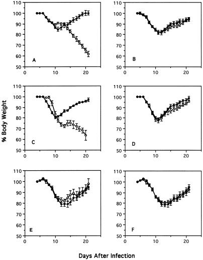
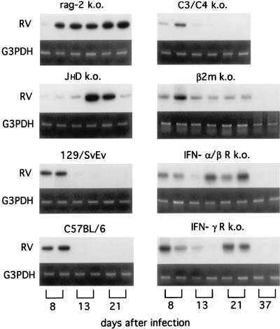
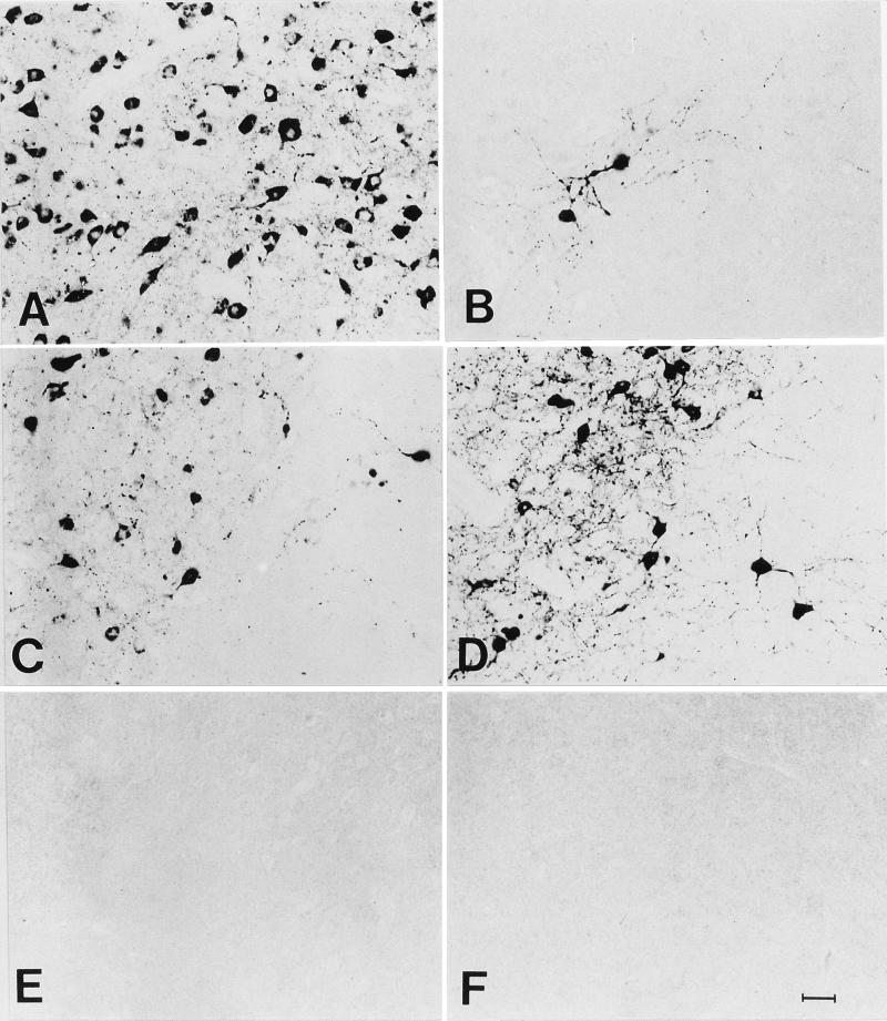
To investigate the involvement of various cellular and humoral aspects of immunity in the clearance of rabies virus from the central nervous system, (CNS), we studied the development of clinical signs and virus clearance from the CNS in knockout mice lacking either B and T cells, CD8+ cytotoxic T cells, B cells, alpha/beta interferon (IFN-alpha/beta) receptors, IFN-gamma receptors, or complement components C3 and C4. Following intranasal infection with the attenuated rabies virus CVS-F3, normal adult mice of different genetic backgrounds developed a transient disease characterized by loss of body weight and appetite depression which peaked at 13 days postinfection (p.i.). While these animals had completely recovered by day 21 p.i., mice lacking either B and T cells or B cells alone developed a progressive disease and succumbed to infection. Mice lacking either CD8+ T cells, IFN receptors, or complement components C3 and C4 showed no significant differences in the development of clinical signs by comparison with intact counterparts having the same genetic background. However, while infectious virus and viral RNA could be detected in normal control mice only until day 8 p.i., in all of the gene knockout mice studied except those lacking C3 and C4, virus infection persisted through day 21 p.i. Analysis of rabies virus-specific antibody production together with histological assessment of brain inflammation in infected animals revealed that clearance of CVS-F3 by 21 days p.i. correlated with both a strong inflammatory response in the CNS early in the infection (day 8 p.i.), and the rapid (day 10 p.i.) production of significant levels of virus-neutralizing antibody (VNA). These studies confirm that rabies VNA is an absolute requirement for clearance of an established rabies virus infection. However, for the latter to occur in a timely fashion, collaboration between VNA and inflammatory mechanisms is necessary.


Figures

FIG. 1
FIG. 1
Effect of i.n. infection with CVS-F3 on body weight of mice with different gene defects. Normal 129/SvEv (A, E, and F) and C57BL/6J (B to D) mice (⧫) and the gene k.o. mice (○), rag-2 k.o. (A), β2m k.o. (B), JHD k.o. (C) C3/C4 k.o. (D), IFN-α/βR k.o. (E), and IFN-γR k.o. (F), were infected i.n. with CVS-F3 as described in Materials and Methods. Mice were weighed on a daily basis. Individual body weights were transformed into percentages, taking the weight at day 5 p.i. as 100%. The results are expressed as mean plus standard error of the mean percent body weight of 15 mice per group.
FIG. 2
FIG. 2
RT-PCR analysis of rabies virus genomic RNA in brain tissue from mice infected i.n. with CVS-F3. Brains from two mice of each of the indicated strains were collected at 8, 13, 21, and 37 days after i.n. infection with CVS-F3, and RNA was extracted and subjected to RT-PCR analysis for rabies virus genomic RNA (RV) as described in Materials and Methods. Amplification of G3PDH mRNA served as an internal control.
FIG. 3
FIG. 3
Immunohistochemical analysis for rabies virus N protein in coronal sections through the hippocampus of rag-2 k.o. (A to C), β2m k.o. (D to F), JHD k.o. (G to I), IFN-γR k.o. (K to M), C57BL/6J (N to P), and 129/SvEv (R and S) mice infected with CVS-F3. Also shown is a section from a noninfected 129/SvEv mouse (Q). Mice were sacrificed at 8 (A, D, G, K, and N), 13 (B, E, H, L, O, and R), and 21 (C, F, I, M, P, and S) days p.i., and brain sections were prepared and stained with antibodies specific for rabies virus N protein as described in Materials and Methods. Arrows indicate N-protein-positive cells. Areas shown at higher magnification in Fig. 4 are marked by asterisks. Bar = 500 μm.
FIG. 4
FIG. 4
Immunohistochemical analysis for rabies virus N protein in coronal sections through the hippocampus of rag-2 k.o. (A), β2m k.o. (B), JHD k.o. (C), IFN-γ R k.o. (D), C57BL/6J (E), and 129/SvEv (F) mice 21 days following infection with CVS-F3. Areas from sections marked by asterisks in Fig. 3 are shown at higher magnification (bar = 25 μm).
FIG. 5
FIG. 5
Histological analysis of coronal sections through the hippocampus of rag-2 k.o. (A to C), β2m k.o. (D to F), JHD k.o. (G to I), IFN-γR k.o. (K to M), C57BL/6J (N to P), and 129/SvEv (R and S) mice infected with CVS-F3. Also shown is a section from a noninfected 129/SvEv mouse (Q). Mice were sacrificed at 8 (A, D, G, K, and N), 13 (B, E, H, L, O, and R), and 21 (C, F, I, M, P, and S) days p.i., and brain sections were prepared and stained with hematoxylin-eosin as described in Materials and Methods. Panels show matching areas of the hippocampal fissure of different mice, magnified approximately 40-fold. Bar = 25 μm.

Similar articles

Cited by

References

    1. Davis D R, Metzger H. Structural basis of antibody function. Annu Rev Immunol. 1983;1:87–117. - PubMed
    1. Dietzschold B. Antibody-mediated clearance of viruses from the mammalian nervous system. Trends Microbiol. 1993;1:63–66. - PMC - PubMed
    1. Dietzschold B, Kao M, Zheng Y M, Chen Z Y, Maul G, Fu Z F, Rupprecht C, Koprowski H. Delineation of putative mechanisms involved in the antibody-mediated clearance of rabies virus from the central nervous system. Proc Natl Acad Sci USA. 1992;89:7252–7256. - PMC - PubMed
    1. Dietzschold B, Schwaeble W, Schaeffer M K-H, Hooper D C, Zheng Y M, Petry F, Sheng H, Fink T, Loos M, Koprowski H, Weihe E. Expression of C1q, a subcomponent of the rat complement system, is dramatically enhanced in brains of rats with either Borna disease or experimental allergic encephalomyelitis. J Neurol Sci. 1995;130:11–16. - PubMed
    1. Dietzschold B, Wiktor T J, Trojanowski J R, Macfarlan R I, Wunner W H, Torres-Anjel M J, Koprowski H. Differences in cell-to-cell spread of pathogenic and apathogenic rabies virus in vivo and in vitro. J Virol. 1985;56:12–18. - PMC - PubMed

Publication types

MeSH terms