. 1998 May;72(5):3762-72.
doi: 10.1128/JVI.72.5.3762-3772.1998.

Transgenic mice secreting coronavirus neutralizing antibodies into the milk

Affiliations

Transgenic mice secreting coronavirus neutralizing antibodies into the milk

I Sola et al. J Virol. 1998 May.

Abstract

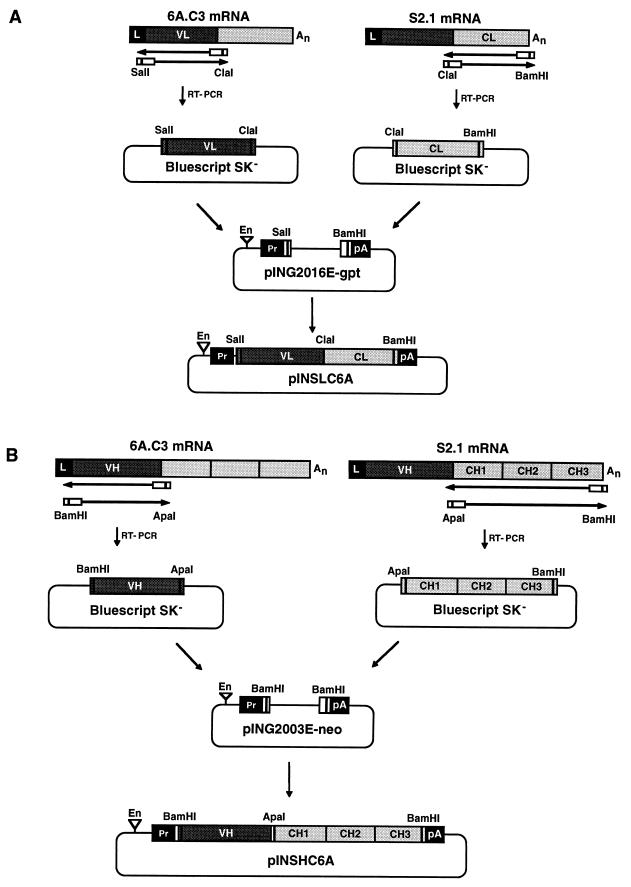
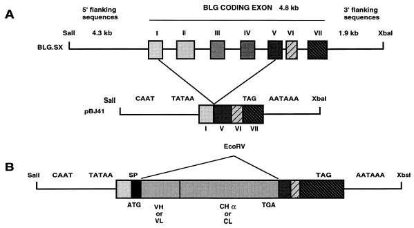
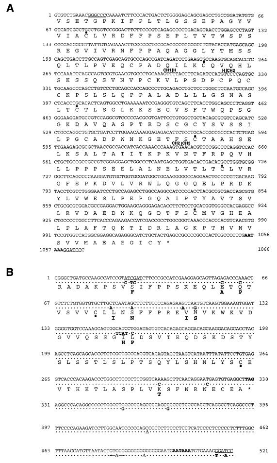
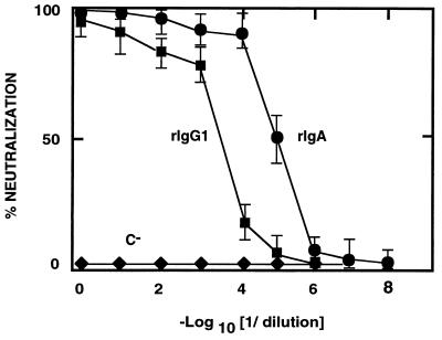
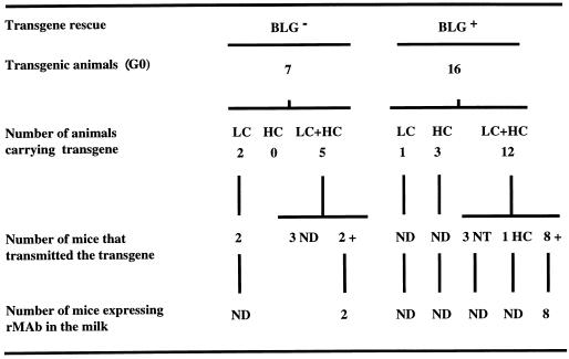
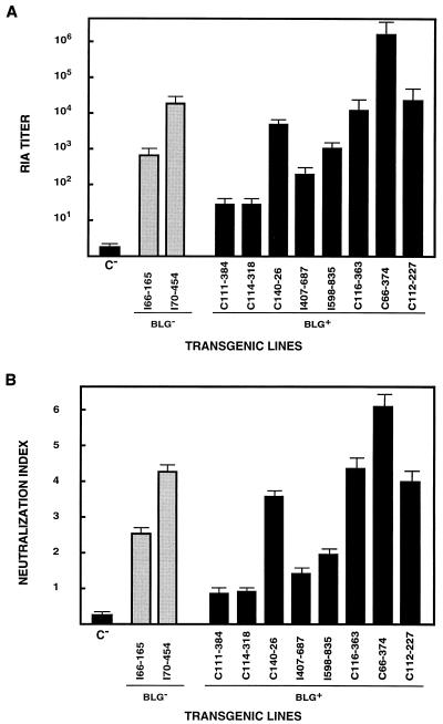
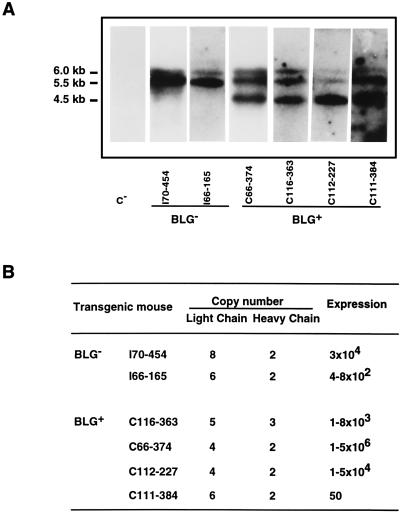
Ten lines of transgenic mice secreting transmissible gastroenteritis coronavirus (TGEV) neutralizing recombinant monoclonal antibodies (rMAbs) into the milk were generated. The rMAb light- and heavy-chain genes were assembled by fusing the genes encoding the variable modules of the murine MAb 6A.C3, which binds an interspecies conserved coronavirus epitope essential for virus infectivity, and a constant module from a porcine myeloma with the immunoglobulin A (IgA) isotype. The chimeric antibody led to dimer formation in the presence of J chain. The neutralization specific activity of the recombinant antibody produced in transiently or stably transformed cells was 50-fold higher than that of a monomeric rMAb with the IgG1 isotype and an identical binding site. This rMAb had titers of up to 10(4) by radioimmunoassay (RIA) and neutralized virus infectivity up to 10(4)-fold. Of 23 transgenic mice, 17 integrated both light and heavy chains, and at least 10 of them transmitted both genes to the progeny, leading to 100% of animals secreting functional TGEV neutralizing antibody during lactation. Selected mice produced milk with TGEV-specific antibody titers higher than 10(6) as determined by RIA, neutralized virus infectivity by 10(6)-fold, and produced up to 6 mg of antibody per ml. Antibody expression levels were transgene copy number independent and integration site dependent. Comicroinjection of the genomic beta-lactoglobulin gene with rMAb light- and heavy-chain genes led to the generation of transgenic mice carrying the three transgenes. The highest antibody titers were produced by transgenic mice that had integrated the antibody and beta-lactoglobulin genes, although the number of transgenic animals generated does not allow a definitive conclusion on the enhancing effect of beta-lactoglobulin cointegration. This approach may lead to the generation of transgenic animals providing lactogenic immunity to their progeny against enteric pathogens.


Figures

FIG. 1
FIG. 1
Cloning of Ig L and H chain cDNAs into expression vectors. (A) Cloning of the recombinant mouse-porcine L-chain cDNA. Poly(A)+ RNA from hybridoma cells secreting MAb 6A.C3 was used as the template for an RT-PCR to obtain the cDNA encoding the VL module. SalI and ClaI restriction sites were introduced into VL module cDNA at the 5′ and 3′ ends, respectively, to facilitate the cloning of the RT-PCR-derived cDNA into pBluescript SK. Poly(A)+ RNA from porcine hybridoma cells secreting MAb S2.1 of the IgA isotype was used as the template for an RT-PCR to obtain the cDNA encoding the CL module. ClaI and BamHI restriction sites were introduced into CL module cDNA at the 5′ and 3′ ends, respectively, to facilitate the cloning of the RT-PCR derived cDNA into pBluescript SK. The resulting VL and CL fragments were joined at the ClaI restriction site and cloned into the expression vector pING2016E-gpt by using the SalI and BamHI sites, yielding plasmid pINSLC6A. (B) Cloning of the recombinant mouse-porcine H-chain cDNA. Poly(A)+ RNA from the hybridoma secreting MAb 6A.C3 was used as the template for an RT-PCR to obtain the cDNA encoding the VH module. BamHI and ApaI restriction sites were introduced into VH module cDNA at the 5′ and 3′ ends, respectively, to facilitate the cloning of the RT-PCR cDNA into pBluescript SK. Poly(A)+ RNA from porcine hybridoma cells secreting MAb S2.1 of the IgA isotype was used as the template for an RT-PCR to obtain the cDNA encoding the CH module. ApaI and BamHI restriction sites were introduced into CH module cDNA at the 5′ and 3′ ends, respectively, to facilitate the cloning of the RT-PCR-derived cDNA into pBluescript SK. The resulting VH and CH fragments were joined at the ApaI restriction site and cloned into the BamHI restriction site of plasmid pING2003E-neo, yielding pINSHC6A. En, SV40 enhancer; Pr, SV40 promoter; An, poly(A) sequence; pA, SV40 polyadenylation signal.
FIG. 2
FIG. 2
Transgene constructs. (A) Structure of the BLG gene (BLG.SX) indicating the introns deleted to generate the pBJ41 plasmid carrying the intronless BLG minigene. The ATG present in the BLG gene was removed in pBJ41, and an EcoRV cloning site was created by introducing a linker between exons I and V. Exon VII does not have an open reading frame. CAAT, TATAA, and AATAAA regulatory signals are indicated. Bold lines indicate 5′- and 3′-flanking sequences and intronic sequences of BLG; boxes indicate BLG exon sequences. (B) Grey boxes, VH- or VL- and CHα- or CL-encoding cDNA segments. All constructs comprise identical 5′- and 3′-regulatory sequences. Relevant restriction enzyme cutting sites are shown. SP, signal peptide.
FIG. 3
FIG. 3
C domain sequences of porcine κ L-chain and α H-chain cDNAs. The sequences of the C Ig domain starting at nucleotide 1 are shown. (A) The nucleotide sequence of the porcine CHα cDNA domain and the deduced amino acid sequence are shown in the first and second lines, respectively. The ApaI and BamHI cloning sites introduced at the 5′ and 3′ ends, respectively, are underlined. Boundaries between domains are indicated by vertical lines and the name of the domain. (B) The nucleotide sequence of the porcine κ L-chain cDNA cloned in our laboratory and the deduced amino acid sequence are shown in the first and third lines, respectively. In the second and fourth lines, the nucleotide and amino acid substitutions in the sequences previously reported (29) for the porcine κ chain are indicated in boldface type. ClaI and BamHI cloning sites introduced at the 5′ and 3′ ends, respectively, are underlined. Nucleotides are numbered at both sides of each line. ▪, cysteine residues predicted to participate in intradomain disulfide bonds. The polyadenylation signal and the stop codons are shown in boldface type. ▵, absent nucleotides.
FIG. 4
FIG. 4
Physical characterization of rMAbs. (A and B) Western blot analysis on 5 to 20% linear gradient SDS-PAGE gels under nonreducing (A) and reducing (B) conditions, developed with rabbit anti-swine IgA and rabbit anti-mouse antisera. (C) Western blot analysis performed under reducing conditions developed with rabbit anti-J-chain antiserum. rIgA, recombinant mouse-swine IgA produced by transformed Sp2/0 cells. rIgG, recombinant mouse-human IgG. IgG, MAb 6A.C3 secreted by the original hybridoma cells. IgM, control polymeric IgM. The positions of the molecular mass markers expressed in kilodaltons are indicated on the left.
FIG. 5
FIG. 5
Comparison of the specific activity of the recombinant antibodies in TGEV neutralization. The neutralization of TGEV by chimeric rIgG1 and rIgA antibodies with the same RIA titer is shown. C, supernatant from myeloma Sp2/0 cells. •, rIgA; ▪, rIgG1; ⧫, negative control MAb. The mean and the standard deviation of three experiments is shown.
FIG. 6
FIG. 6
Transmission and expression of transgenes in BLG and BLG+ transgenic mice. Transgene integration was screened in tail DNA samples from G0 and G1 mice by PCR with primers detecting the constant L (LC), constant H (HC), and BLG transgenes. PCR-positive samples were further analyzed by Southern blotting with BLG-specific probes. In several lines, analysis was extended to the G2 and G3 generation of mice. Transgene expression in milk was determined by RIA with G1 females. ND, not determined; NT, no Ig chain transmission; HC, mice which transmitted only the constant H chain.
FIG. 7
FIG. 7
Recombinant IgA expression in the milk of transgenic mice. (A) The RIA titers for rIgA in the milk of BLG and BLG+ transgenic mice are shown as the mean of the antibody titers in three to six milk samples from the same transgenic mouse, collected at different days through lactation. C, milk from nontransgenic mice. (B) Neutralization index of rIgA present in the milk of BLG and BLG+ transgenic lines. The neutralization assays were performed with same milk samples used for the experiment in panel A.
FIG. 8
FIG. 8
rIgA expression in the milk from transgenic mice during lactation. (A) The titers of rIgA in the milk of transgenic mice I70-454 and C66-374, generated by microinjecting the Ig chains alone (BLG) or with the BLG gene (BLG+), during a lactation cycle are represented. These transgenic mice were chosen because their milk had the highest RIA titers. C, milk from a nontransgenic mouse. (B) Neutralization index for rIgA in the milk of I70-454 (BLG) and C66-374 (BLG+) transgenic mice during lactation.
FIG. 9
FIG. 9
Relationship between copy number and rIgA expression in the milk. (A) Southern blot analysis of BLG and BLG+ transgenic lines. C, nontransgenic mouse DNA. Tail DNA samples were cleaved with EcoRI and analyzed by electrophoresis and Southern blotting. Filters were probed with 5′-flanking sequences common to the three transgenes. The 6.0-kb BLG-SHC, 5.5-kb BLG-SLC, and 4.5-kb BLG transgene-specific EcoRI fragments are shown. (B) Comparison of rIgA titers in BLG and BLG+ transgenic lines carrying different transgene copy numbers. The expression levels are reported as the RIA titers. The RIA titers of rIgA in the milk of BLG and BLG+ transgenic mice were calculated as the mean of the antibody titers in three to six milk samples from the same mouse.

References

    1. Ali S, Clark A J. Characterization of the gene encoding ovine β-lactoglobulin. J Mol Biol. 1988;199:415–426. - PubMed
    1. Ali S, McClenaghan M, Simons J P, Clark A J. Characterisation of the alleles encoding ovine β-lactoglobulins A and B. Gene. 1990;91:201–207. - PubMed
    1. Antón I M, Suñé C, Meloen R H, Borrás-Cuesta F, Enjuanes L. A transmissible gastroenteritis coronavirus nucleoprotein epitope elicits T helper cells that collaborate in the in vitro antibody synthesis to the three major structural viral proteins. Virology. 1995;212:746–751. - PMC - PubMed
    1. Armstrong S J, Outlaw M C, Dimmock N J. Morphological studies of neutralization of influenza virus by IgM. J Gen Virol. 1990;71:2313–2319. - PubMed
    1. Brim T A, VanCott J L, Lunney J K, Saif L J. Lymphocyte proliferation responses of pigs inoculated with transmissible gastroenteritis virus or porcine respiratory coronavirus. Am J Vet Res. 1994;55:494–501. - PubMed

Publication types

MeSH terms

LinkOut - more resources