. 1998 May;72(5):3819-26.
doi: 10.1128/JVI.72.5.3819-3826.1998.

Two RING finger proteins, the oncoprotein PML and the arenavirus Z protein, colocalize with the nuclear fraction of the ribosomal P proteins

Affiliations

Two RING finger proteins, the oncoprotein PML and the arenavirus Z protein, colocalize with the nuclear fraction of the ribosomal P proteins

K L Borden et al. J Virol. 1998 May.

Abstract

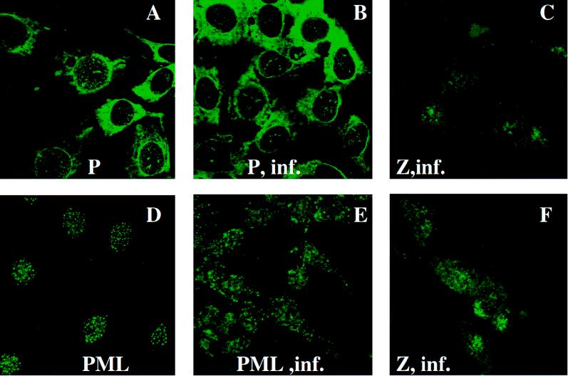
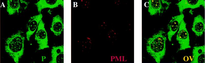
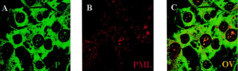
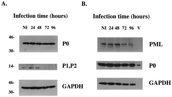
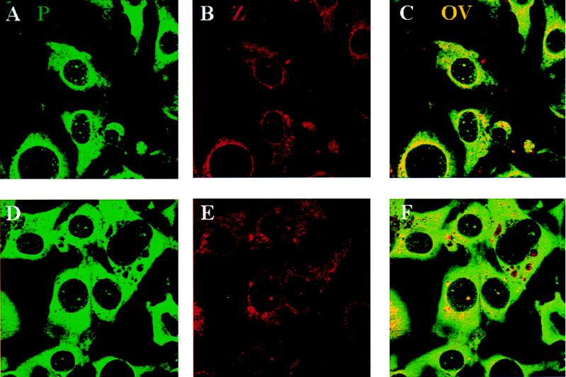
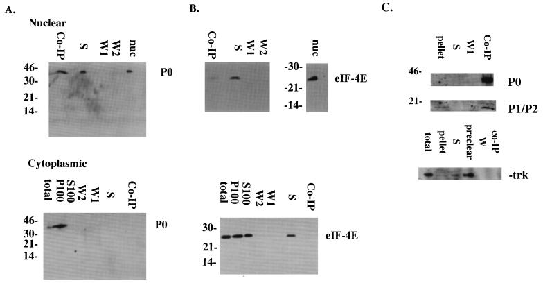
The promyelocytic leukemia (PML) protein forms nuclear bodies which are relocated to the cytoplasm by the RNA virus lymphocytic choriomeningitis virus (LCMV). The viral Z protein directly binds to PML and can relocate the nuclear bodies. Others have observed that LCMV virions may contain ribosomes; hence, we investigated the effects of infection on the distribution of ribosomal P proteins (P0, P1, and P2) with PML as a reference point. We demonstrate an association of PML bodies with P proteins by indirect immunofluorescence and coimmunoprecipitation experiments, providing the first evidence of nucleic acid-binding proteins associated with PML bodies. We show that unlike PML, the P proteins are not redistributed upon infection. Immunofluorescence and coimmunoprecipitation studies indicate that the viral Z protein binds the nuclear, but not the cytoplasmic, fraction of P0. The nuclear fraction of P0 has been associated with translationally coupled DNA excision repair and with nonspecific endonuclease activity; thus, P0 may be involved in nucleic acid processing activities necessary for LCMV replication. During the infection process, PML, P1, and P2 are downregulated but P0 remains unchanged. Further, P0 is present in virions while PML is not, indicating some selectivity in the assembly of LCMV.


Figures

FIG. 1
FIG. 1
Effect of LCMV infection on P proteins, PML, and Z in NIH 3T3 cells. Experiments were carried out as described in Materials and Methods. A and B, Cells stained with anti-P protein antibody in uninfected and infected (inf.) cells (90 h), respectively; D and E, Cells stained with the PML polyclonal antibody in uninfected and infected cells (90 h), respectively; C and F, staining for affinity-purified Z antibody in cells infected for 90 h. Magnifications: ×100 for A and B and ×40 for C to F with zooms of 3.3 (C), 2.6 (D), 2.8 (E), and 3.2 (F). Panels A and B represent single sections through the cell, whereas C, D, E, and F represent projections of several sections through the entire cell.
FIG. 2
FIG. 2
PML and P proteins colocalize in uninfected NIH 3T3 cells. Panels: A, cells stained with the P protein antibody (green); B, cells stained with the PML antibody (red); C, overlay (OV) (yellow). These confocal micrographs represent single slices through the plane of cells. Magnification, ×100. FITC was excited at 488 nm, and Texas Red was excited at 568 nm. The two channels were recorded independently.
FIG. 3
FIG. 3
PML and P proteins colocalize in NIH 3T3 cells infected for 90 h with LCMV. Panels: A, cells stained with the P protein antibody (green); B, cells stained with the PML antibody (red); C, overlay (OV) (yellow). These confocal micrographs represent single slices through the plane of cells. Magnification, ×100. FITC was excited at 488 nm, and Texas Red was excited at 568 nm. The two channels were recorded independently.
FIG. 4
FIG. 4
The PML, P1, and P2 proteins are downregulated during infection, but the levels of P0 remain unchanged. Infection and Western blot analysis were carried out as described in Materials and Methods. NI, not infected. The levels of GAPDH were measured during the same time course as a control. In panel B, blots were prepared with virion preparations (lane V), as well as infected cells, as in panel A. The values to the left are molecular sizes in kilodaltons.
FIG. 5
FIG. 5
The viral Z protein and the P proteins colocalize in infected cells. Cells were infected for 90 h. Panels A and D show cells stained with the P protein antibody (green), panels B and E show cells stained with the affinity-purified Z antibody (red), and panels C and F show the overlay (OV) (yellow). These confocal micrographs represent single slices through the plane of cells. Magnification, ×100. FITC was excited at 488 nm, and Texas Red was excited at 568 nm. The two channels were recorded independently.
FIG. 6
FIG. 6
Direct interaction between the Z and P proteins in transfected cells (A). Cells were fractionated and coimmunoprecipitated with the Z antibody as described in Materials and Methods. In panel B, blots were probed with a control antibody, eIF-4E (140 kDa) to indicate that the coimmunoprecipitations were specific (see text for details). In panel C, cell lysates were coimmunoprecipitated with MAb 5E10 and probed with the P protein antibody as described in Materials and Methods. As a negative control, blots were probed with the trk antibody. Lanes: Co-IP, coimmunoprecipitated fraction; S, supernatant after coimmunoprecipitation; W1 and W2, washes one and two, respectively, after coimmunoprecipitation; nuc, nuclear fraction; total, total cell lysate. P100 and S100 are described in Materials and Methods. The values on the left are molecular sizes in kilodaltons.

References

    1. Ascoli C A, Maul G G. Identification of a novel nuclear domain. J Cell Biol. 1991;112:785. - PMC - PubMed
    1. Boddy M N, Howe K, Etkin L D, Solomon E, Freemont P S. PIC1, a novel ubiquitin-like protein which interacts with the PML component of a multiprotein complex that is disrupted in acute promyelocytic leukaemia. Oncogene. 1996;13:971–982. - PubMed
    1. Borden K L B, CampbellDwyer E J, Salvato M S. An arenavirus RING (zinc-binding) protein binds the oncoprotein PML and relocates PML nuclear bodies to the cytoplasm. J Virol. 1998;72:758–766. - PMC - PubMed
    1. Borden K L B, CampbellDwyer E J, Salvato M S. The promyelocytic leukemia protein PML has a pro-apoptotic activity mediated through its RING. FEBS Lett. 1997;418:30–34. - PMC - PubMed
    1. Borden K L B, Freemont P S. The RING finger: an example of a sequence structure family. Curr Opin Struct Biol. 1996;6:395–401. - PubMed

Publication types

MeSH terms

LinkOut - more resources