. 1998 May;72(5):3827-36.
doi: 10.1128/JVI.72.5.3827-3836.1998.

Hepatitis C virus structural proteins assemble into viruslike particles in insect cells

Affiliations

Hepatitis C virus structural proteins assemble into viruslike particles in insect cells

T F Baumert et al. J Virol. 1998 May.

Abstract

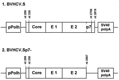
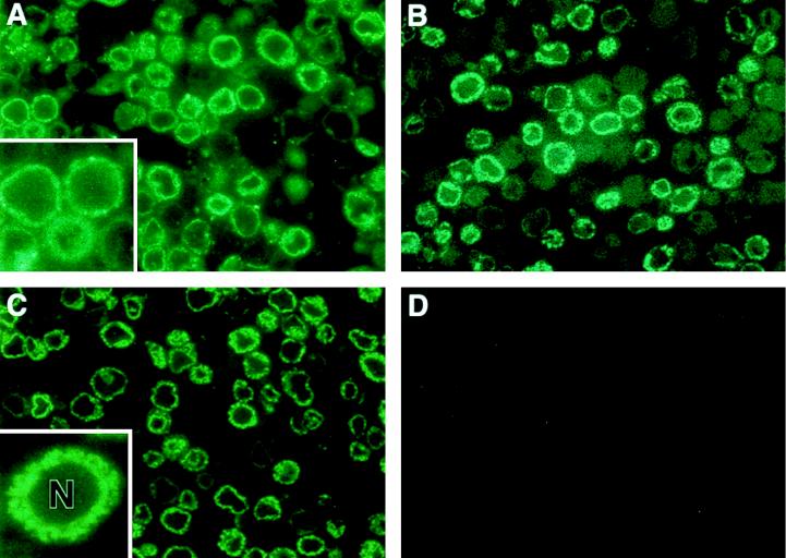
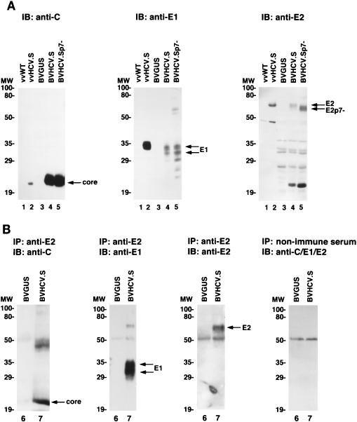
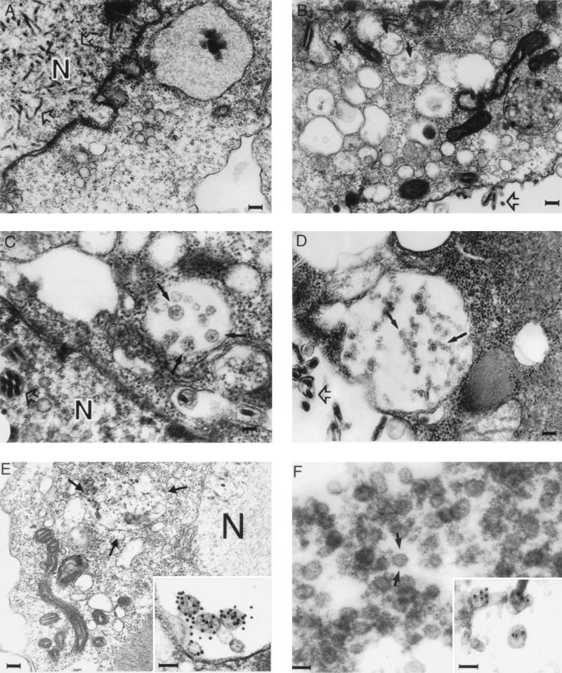
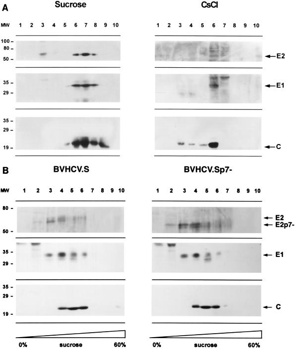
Hepatitis C virus (HCV) is a leading cause of chronic hepatitis in the world. The study of HCV has been hampered by the low level of viral particles in infected individuals, the inability to propagate efficiently the virus in cultured cells, and the lack of a convenient animal model. Due to these obstacles, neither the structure of the virus nor the prerequisites for its assembly have been clearly defined. In this report, we describe a model for the production and purification of HCV-like particles in insect cells using a recombinant baculovirus containing the cDNA of the HCV structural proteins. In insect cells, expressed HCV structural proteins assembled into enveloped viruslike particles (40 to 60 nm in diameter) in large cytoplasmic cisternae, presumably derived from the endoplasmic reticulum. Biophysical characterization of viruslike particles by CsCl and sucrose gradient centrifugation revealed biophysical properties similar to those of putative virions isolated from infected humans. The results suggested that HCV core and envelope proteins without p7 were sufficient for viral particle formation. Analysis of particle-associated nucleic acids demonstrated that HCV RNAs were selectively incorporated into the particles over non-HCV transcripts. The synthesis of HCV-like particles in insect cells may provide an important tool to determine the structural requirements for HCV particle assembly as well as to study viral genome encapsidation and virus-host interactions. The described system may also represent a potential approach toward vaccine development.


Figures

FIG. 1
FIG. 1
Map depicting segments of the HCV genome in the recombinant baculoviruses expressing HCV structural proteins. The BVHCV.S construct contains a short stretch of the 5′ UTR as well as the coding sequences for the core, E1, E2, and p7 proteins and 21 amino acids of the NS2 protein. The polyprotein open reading frame begins at nt 330, and the junction of E2-p7 and NS2 is at nt 2756. BVHCV.Sp7− contains the same 5′ UTR, core, E1, and E2 coding sequences as BVHCV.S but not p7 and the amino-terminal part of the NS2 protein. pPolh, baculovirus polyhedrin promoter; SV40, simian virus 40.
FIG. 2
FIG. 2
Immunofluorescence of HCV structural proteins expressed in insect cells. (A, B, and C) Insect cells were infected with the recombinant baculovirus BVHCV.S containing the cDNA for the HCV structural proteins at an MOI of 10. At 96 h postinfection, the cells were fixed and processed as described in Materials and Methods. Protein expression was analyzed with serum obtained from an HCV-infected human (A), anti-E1 antibody (B), or anti-E2 antibody (C) as described in Materials and Methods. The insets show a higher magnification of the stained insect cells. Immunostaining was confined to the cytosol, whereas the nucleus (N) remained unstained. (D) Insect cells infected with the control baculovirus BVGUS containing the cDNA for GUS were subjected to staining with a mixture of the same serum and antibodies as those shown in panels A, B, and C.
FIG. 3
FIG. 3
(A) Expression of HCV structural proteins in insect and mammalian cells. Sf9 insect cells were infected either with control baculovirus BVGUS (lane 3) or with the HCV expression baculoviruses BVHCV.S (lane 4) and BVHCV.Sp7− (lane 5). At 96 h postinfection, the cells were lysed as described in Materials and Methods. To compare the expression of HCV structural proteins between insect and mammalian cells, BSC-1 cells were infected with a wild-type vaccinia virus (vvWT) (lane 1) or a vaccinia virus (vvHCV.S) containing the same cDNA for the HCV structural proteins as baculovirus BVHCV.S (lane 2). At 8 h postinfection, the cells were lysed as described in Materials and Methods. Sf9 and BSC-1 cell lysates were then subjected to SDS-PAGE and immunoblotting (IB) with anti-core (left panel), anti-E1 (middle panel), or anti-E2 (right panel) antibodies. Molecular masses (in kilodaltons) of protein molecular weight (MW) markers are indicated on the left; HCV-specific proteins are indicated on the right. (B) Coimmunoprecipitation of HCV structural proteins in insect cells. Insect cells were infected with BVGUS (lane 6) or BVHCV.S (lane 7). At 96 h postinfection, the cells were lysed and subjected to immunoprecipitation (IP) with an anti-E2 antibody or nonimmune serum as indicated. The immunoprecipitated proteins were subjected to SDS-PAGE and immunoblotting (IB) with anti-core (C), anti-E1, or anti-E2 antibodies as indicated; the control immunoprecipitate with nonimmune serum was probed with a mixture of antibodies to core, E1, and E2 proteins. The exposure times for the probed blot were adjusted to visualize the proteins and do not represent a quantitation of the precipitated proteins.
FIG. 4
FIG. 4
Electron microscopy of HCV-like particles in insect cells infected with BVHCV.S or BVHCV.Sp7−. Insect cells in monolayer cultures were infected with BVGUS, BVHCV.S, or BVHCV.Sp7−. At 96 h postinfection, the cells were fixed and processed for electron microscopy as described in Materials and Methods. (A) Insect cells infected with the control baculovirus BVGUS. Electron microscopy demonstrates abundant replicative forms of baculovirus (open arrows) in the nucleus (N). No HCV-like particles can be seen in the cytoplasm. Bar, 120 nm. (B) Insect cells infected with BVHCV.S. Electron microscopy demonstrates numerous enveloped, viruslike particles 40 to 60 nm in diameter (closed arrows) in vacuoles. In addition, the synthesis of baculovirus is visualized (open arrow). Bar, 120 nm. (C) Higher magnification of viruslike particles (closed arrows) seen in BVHCV.S-infected cells. Bar, 40 nm. (D) Insect cells infected with BVHCV.Sp7−. Electron microscopy demonstrates viruslike particle formation in a large cytoplasmic vacuole similar to that seen in BVHCV.S-infected cells (solid arrows). Baculovirus particles (open arrows) can be distinguished easily from the HCV-like particles in both panels C and D. Bar, 100 nm. (E) Immunogold labeling of viruslike particles (arrows) similar to those seen in panels B, C, and D with serum from an HCV-infected individual. Immunostaining was confined to the cytoplasmic cisternae and ER, whereas the N remained unstained. Bar, 120 nm. The inset shows a higher magnification of the viruslike particles labeled with an anti-E2 antibody. Bar, 50 nm. (F) Electron microscopy of viruslike particles (arrows) partially purified by sucrose gradient centrifugation. Bar, 50 nm. The inset shows labeling of partially purified particles with an anti-E2 antibody. Bar, 50 nm.
FIG. 5
FIG. 5
Purification of HCV-like particles by sucrose and CsCl gradient centrifugation. (A) Sucrose and CsCl equilibrium centrifugation. At 96 h postinfection, insect cells infected with BVHCV.S were lysed and subjected to low-speed centrifugation, and the supernatant was pelleted over a 30% sucrose cushion. The pellet containing the viruslike particles was resuspended and subjected to a second sucrose or CsCl equilibrium gradient centrifugation as described in Materials and Methods. Ten fractions were collected from the top and analyzed by SDS-PAGE and immunoblotting with anti-core (C), anti-E1, or anti-E2 antibodies. Molecular masses (in kilodaltons) of protein molecular weight (MW) markers are indicated on the left; HCV-specific proteins are indicated on the right. (B) Sucrose sedimentation velocity centrifugation. Lysates of insect cells infected with BVHCV.S and BVHCV.Sp7− were layered onto a 10 to 60% sucrose gradient and centrifuged for 2.5 h at 4°C and 200,000 × g. Ten fractions were collected from the top and analyzed for HCV structural proteins as described above.
FIG. 6
FIG. 6
(A) Analysis of total RNA of insect cells infected with BVHCV.S, BVGUS, or a baculovirus containing the cDNA for the HIV glycoprotein precursor gp160 (BVHIV). At 96 h postinfection, total RNA was isolated as described in Materials and Methods and subjected to Northern blot analysis with an HCV (lane 1)-, GUS (lane 2)-, or HIV gp160 (lane 3)-specific cDNA probe. Ethidium bromide staining of the 18S RNA was used as a control for RNA loading. (B and C) Encapsidation of HCV RNA into HCV-like particles. In order to distinguish between preferential encapsidation and random, nonspecific incorporation of HCV RNA by the viruslike particles, insect cells were coinfected with BVHCV.S, BVGUS, and BVHIV. HCV-like particles were isolated by immunoprecipitation (IP) (B) or sucrose gradient centrifugation followed by immunoprecipitation (IP) (C) as described in Materials and Methods. Particle-associated RNA was purified as described in Materials and Methods and analyzed by Northern blotting with an HCV-specific cDNA probe (HCV nt 259 to 2819; upper blots) or a GUS- and HIV gp160-specific cDNA probe (lower blots). IgG, immunoglobulin G.

References

    1. Acs G, Sells M A, Purcell R H, Price P, Engle R, Shapiro M, Popper H. Hepatitis B virus produced by transfected HepG2 cells causes hepatitis in chimpanzees. Proc Natl Acad Sci USA. 1987;84:4641–4644. - PMC - PubMed
    1. Alter H J, Purcell R H, Shih J W, Melpolder J C, Houghton M, Choo Q-L, Kuo G. Detection of antibody to hepatitis C virus in prospectively followed transfusion recipients with acute and chronic non-A, non-B hepatitis. N Engl J Med. 1989;321:1494–1500. - PubMed
    1. Baumert T F, Rogers S A, Hasegawa K, Liang T J. Two core promoter mutations identified in a hepatitis B virus strain associated with fulminant hepatitis result in enhanced viral replication. J Clin Invest. 1996;98:2268–2276. - PMC - PubMed
    1. Bielefeldt Ohmann H, Bloch B. Electron microscopic studies of bovine viral diarrhea virus in tissues of diseased calves and in cell cultures. Arch Virol. 1981;71:57–74. - PubMed
    1. Chomczynski P N, Sacchi N. Single-step method of RNA isolation by acid guanidinium thiocyanate-phenol-chloroform extraction. Anal Biochem. 1987;162:156–159. - PubMed

Publication types

MeSH terms

LinkOut - more resources