. 1998 May;72(5):3872-86.
doi: 10.1128/JVI.72.5.3872-3886.1998.

Simian immunodeficiency virus replicates to high levels in sooty mangabeys without inducing disease

Affiliations

Simian immunodeficiency virus replicates to high levels in sooty mangabeys without inducing disease

M A Rey-Cuillé et al. J Virol. 1998 May.

Abstract

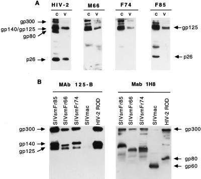
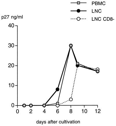
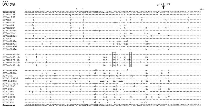
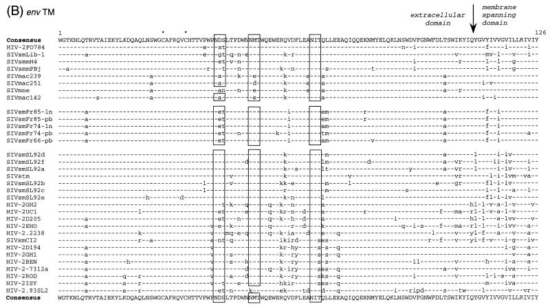
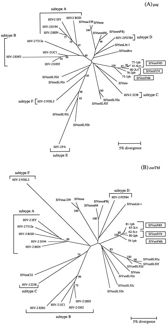
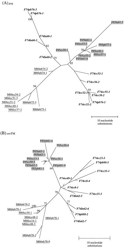
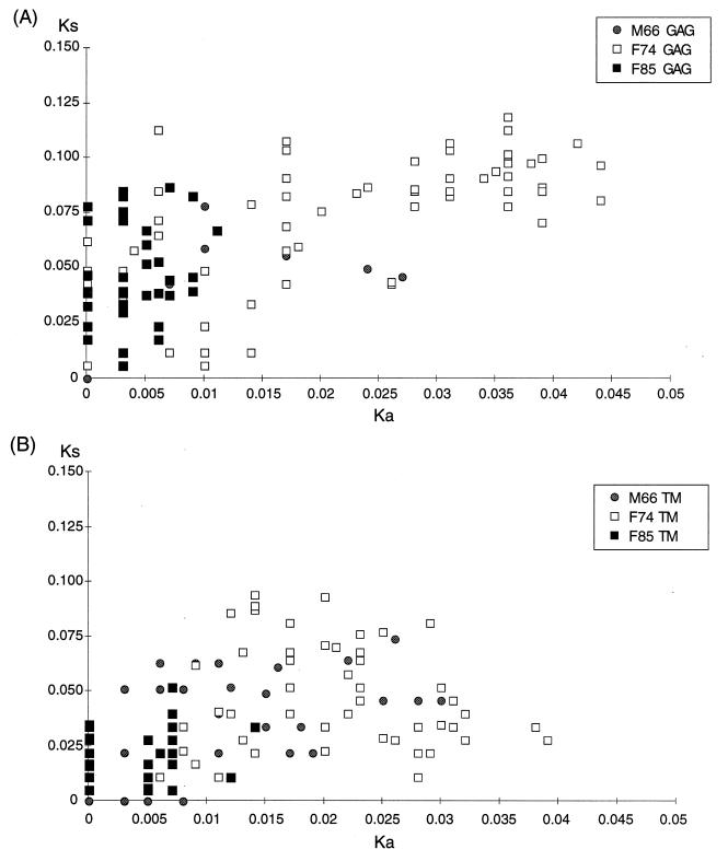
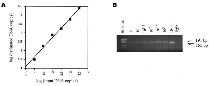
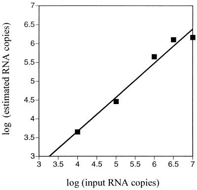
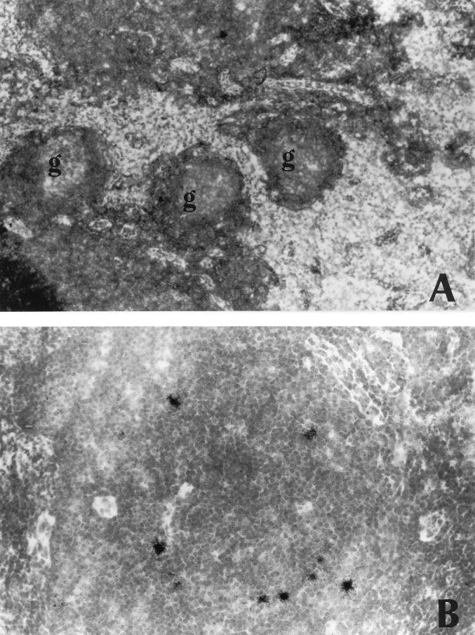
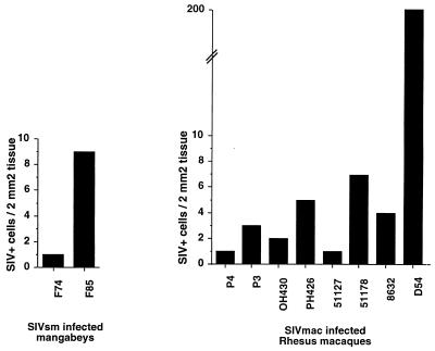
A serologic survey of primates living in a French zoo allowed identification of three cases of infection with simian immunodeficiency virus in sooty mangabeys (Cercocebus atys) (SIVsm). Viral isolates, which were designated SIVsmFr66, SIVsmFr74, and SIVsmFr85, were obtained after short-term culture of mangabey lymphoid cells. Phylogenetic analysis of gag and env sequences amplified directly from mangabey tissues showed that the three SIVsmFr were genetically close and that they constituted a new subtype within the diverse SIVsm-SIVmac-human immunodeficiency virus type 2 (HIV-2) group. We could reconstruct the transmission events that likely occurred in 1986 between the three animals and evaluate the divergence of SIVsmFr sequences since transmission. The estimated rate of mutation fixation was 6 x 10(-3) substitutions per site per year, which was as high as the rate found for SIVmac infection in macaques. These data indicated that SIVsmFr replicated at a high rate in mangabeys, despite the nonpathogenic character of infection in this host. The viral load evaluated by competitive PCR reached 20,000 viral DNA copies per 10(6) lymph node cells. In addition, productively infected cells were readily detected in mangabey lymphoid tissues by in situ hybridization. The amounts of viral RNA in plasma ranged from 10(5) to 10(7) copies per ml. The cell-associated and plasma viral loads were as high as those seen in susceptible hosts (humans or macaques) during the asymptomatic stage of HIV or SIVmac infections. Thus, the lack of pathogenicity of SIVsm for its natural host cannot be explained by limited viral replication or by tight containment of viral production.


Figures

FIG. 1
FIG. 1
Analysis of mangabey serologic reactivity and of SIVsmFr protein profile by Western blotting. (A) Reactivity of a typical human HIV-2-positive serum (left lane) and of three mangabey serum samples (M66, F74, and F85) with HIV-2 ROD antigens. The sera were tested on proteins extracts from HIV-2-infected cells (c) and from HIV-2 viral pellets (v). The human serum reacted with the glycoprotein precursor (gp 140) and its dimer (gp300), the surface glycoprotein (gp125), the dimer of transmembrane glycoproteins (gp80), and the Gag core p27. The mangabey sera reacted mostly with envelope antigens, while their reactivities to Gag p27 were limited or undetectable. (B) Profile of SIVsmFr proteins detected with anti-HIV-2 MAb directed to the surface glycoprotein (125-B) and to the transmembrane glycoprotein (1H8). Protein extracts from CEMX174 cells infected with either of the three SIVsmFr isolates, SIVmac251, or HIV-2 ROD were tested with MAb 125-B (left panel) or with MAb 1H8 (right panel).
FIG. 2
FIG. 2
Isolation of SIVsmFr74 from mangabey PBMC and LNC. Virus isolation was performed by coculturing F74 PBMC, LNC, and LNC from which CD8+ cells were depleted (LNC CD8) with human PBMC in the presence of IL-2 and PHA. Virus production was monitored by an antigen-capture assay specific for SIV p27 Gag.
FIG. 3
FIG. 3
Multiple alignement of amino acid sequences comparing SIVsmFr with other viruses of the HIV-2–SIVsm–SIVmac lineage. Five representative SIVsmFr clones amplified from lymph nodes DNA (ln) or from PBMC DNA (pb) were aligned with previously published sequences (52). (A) Alignment of Gag amino acid sequences. The junction of the matrix p17 with the core protein p27 is indicated by an arrow. A glutamic acid rich sequence unique to SIVsmFr viruses is boxed. (B) Alignment of transmembrane glycoprotein (TM) amino acid sequences. The limits of the extracellular domain with the hydrophobic membrane-spanning domain is indicated by an arrow. Stars, cysteine residues; boxes, conserved glycosylation sites in TM; dashes, sequence identity with the consensus; dots, gaps introduced to optimize the alignment.
FIG. 3
FIG. 3
Multiple alignement of amino acid sequences comparing SIVsmFr with other viruses of the HIV-2–SIVsm–SIVmac lineage. Five representative SIVsmFr clones amplified from lymph nodes DNA (ln) or from PBMC DNA (pb) were aligned with previously published sequences (52). (A) Alignment of Gag amino acid sequences. The junction of the matrix p17 with the core protein p27 is indicated by an arrow. A glutamic acid rich sequence unique to SIVsmFr viruses is boxed. (B) Alignment of transmembrane glycoprotein (TM) amino acid sequences. The limits of the extracellular domain with the hydrophobic membrane-spanning domain is indicated by an arrow. Stars, cysteine residues; boxes, conserved glycosylation sites in TM; dashes, sequence identity with the consensus; dots, gaps introduced to optimize the alignment.
FIG. 4
FIG. 4
Phylogenetic relationships among the newly characterized SIVsmFr and viruses of the HIV-2–SIVsm–SIVmac lineage. (A) Phylogenetic tree derived from gag sequences. The analysis was performed on a 369-bp fragment that encompassed the Gag p17-p26 junction. (B) Phylogenetic tree derived from env sequences. A 380-bp fragment coding for a portion of the extracellular and membrane-spanning domains of TM was analyzed. Phylogenetic trees were constructed by the neighbor-joining method on 1,000 bootstrap replicates of the data. Values on the nodes are percentages of bootstraps with which the cluster is supported; only values greater than 70% are shown. The new SIVsmFr sequences are boxed; HIV-2 subtypes A to F, as defined in references and , are indicated.
FIG. 5
FIG. 5
Phylogenetic relationships among SIVsmFr clones. (A) Phylogenetic tree derived from sequence comparison of gag clones. (B) Phylogenetic tree derived from sequence comparison of env (TM) clones. The trees were constructed by the maximum likelihood method implemented in the FastDNAml program, with 100 bootstrap replicates of the data. Values on the nodes are percentages of bootstraps with which the cluster is supported; only values greater than 70% are shown. SIVsmFr66 clones are underlined, SIVsmFr74 clones are in boldface type, and SIVsmFr85 clones are shaded. Abbreviations within the names of the clones indicate whether they were amplified from PBMC (pb), lymph nodes (ln), or short-term cocultures with human PBMC (cc).
FIG. 6
FIG. 6
Comparison of synonymous and nonsynonymous mutations between pairs of sequences. Ks and Ka values between pairs of clones obtained from the same animal were measured. The range of the Ka parameter was limited for F85, indicating homogeneity of viral amino acid sequences in this animal.
FIG. 7
FIG. 7
Quantitation of SIVsmFr by competitive PCR. (A) Calibration of the competitive PCR assay. Test samples containing known amounts of a SIVsmFr74 gag plasmid were assayed by competitive PCR. The log of the DNA copy number estimated in the assay was plotted as a function of the log of input plasmid copy number. Each plot symbol corresponds to the result of a series of amplifications in the presence of different dilutions of competitor DNA fragment. The competitive PCR assay gave a linear response over the range of concentrations studied (y = 1.10 x + 0.53; r = 0.995). (B) Example of a competitive PCR. Lymph node DNA from animal F85 was amplified in the presence of serial dilutions of competitor DNA. The PCR products were separated by electrophoresis on a 3% agarose gel. The endogenous SIV product can be distinguished from the competitor product by its size, which is larger by 36 bp. m.w.m., molecular weight marker.
FIG. 8
FIG. 8
Calibration of the competitive RT-PCR assay. Known amounts of RNA transcribed from a gag plasmid clone (F74cc55-1) were assayed by competitive RT-PCR. The log of the SIV RNA copy number estimated in the assay was plotted as a function of the log of the input RNA copy number. The competitive RT-PCR assay gave a linear response over the range of concentrations studied (y = 0.91 x + 0.03; r = 0.987).
FIG. 9
FIG. 9
Detection of SIV expression in lymph nodes by in situ hybridization. Frozen lymph node sections were hybridized with a 35S-labeled riboprobe specific for SIVsmFr RNA. (A) Low-magnification view of F85 lymph node. The histology is normal, with follicles of a limited size. No hybridization signal could be detected in the germinal centers (g) (original magnification, ×100); (B) higher-magnification view of F85 lymph node showing numerous infected cells scattered in the paracortical region. The intensity of the hybridization signal indicates that the cells are productively infected (original magnification; ×250).
FIG. 10
FIG. 10
Comparison of the numbers of productively infected cells in mangabey and macaque lymph nodes. The numbers of productively infected cells detected by in situ hybridization in a 2-mm2 area of lymph node section were counted. The mean counts obtained for three sections are indicated. Macaque lymph nodes were analyzed 2 months after inoculation of SIVmac251. Mangabey lymph nodes were obtained from naturally infected animals.

Similar articles

Cited by

References

    1. Ahmed-Ansari A, Powell J D, Jensen P E, Yehuda-Cohen T, McClure H M, Anderson D, Fultz P N, Sell K W. Requirement for simian immunodeficiency virus antigen-specific in vitro proliferation of T-cells from infected rhesus macaques and sooty mangabeys. AIDS. 1990;4:399–407. - PubMed
    1. Alexander L, Du Z, Rosenzweig M, Jung J U, Desrosiers R C. A role for natural simian immunodeficiency virus and human immunodeficiency virus type 1 Nef alleles in lymphocyte activation. J Virol. 1997;71:6094–6099. - PMC - PubMed
    1. Allan J S, Short M, Taylor M E, Su S, Hirsh V M, Johnson P R, Shaw G M, Hahn B H. Species-specific diversity among simian immunodeficiency viruses from African green monkeys. J Virol. 1991;65:2816–2828. - PMC - PubMed
    1. Baier M, Dittmar M T, Cichutek K, Kurth R. Development of in vivo genetic variability of simian immunodeficiency virus. Proc Natl Acad Sci USA. 1991;88:8126–8130. - PMC - PubMed
    1. Balfe P, Simmonds P, Ludlam C A, Bishop J O, Brown A J. Concurrent evolution of human immunodeficiency virus type 1 in patients infected from the same source: rate of sequence change and low frequency of inactivating mutations. J Virol. 1990;64:6221–6233. - PMC - PubMed

Publication types

MeSH terms

LinkOut - more resources