. 1998 May;72(5):3980-90.
doi: 10.1128/JVI.72.5.3980-3990.1998.

Natural isolates of simian virus 40 from immunocompromised monkeys display extensive genetic heterogeneity: new implications for polyomavirus disease

Affiliations

Natural isolates of simian virus 40 from immunocompromised monkeys display extensive genetic heterogeneity: new implications for polyomavirus disease

J A Lednicky et al. J Virol. 1998 May.

Abstract

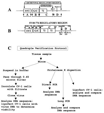
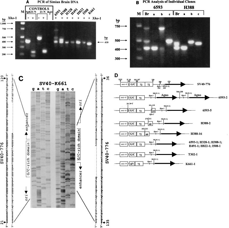
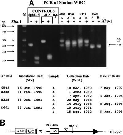
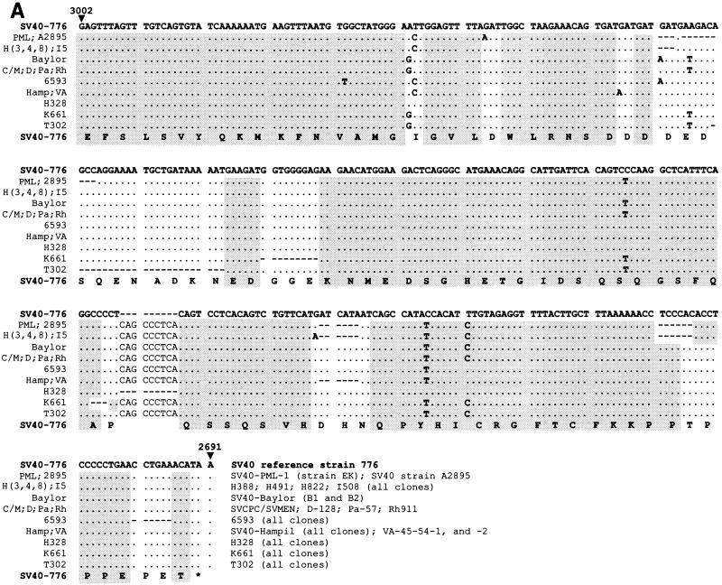
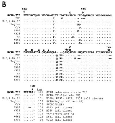
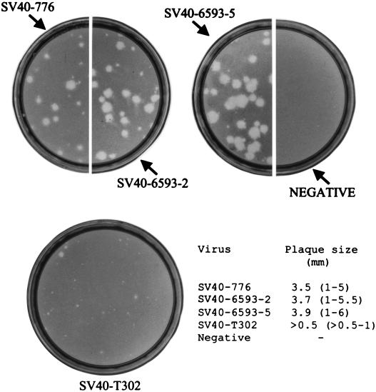
Simian virus 40 (SV40) DNAs in brain tissue and peripheral blood mononuclear cells (PBMCs) of eight simian immunodeficiency virus-infected rhesus monkeys with SV40 brain disease were analyzed. We report the detection, cloning, and identification of five new SV40 strains following a quadruple testing-verification strategy. SV40 genomes with archetypal regulatory regions (containing a duplication within the G/C-rich regulatory region segment and a single 72-bp enhancer element) were recovered from seven animal brains, two tissues of which also contained viral genomes with nonarchetypal regulatory regions (containing a duplication within the G/C-rich regulatory region segment as well as a variable duplication within the enhancer region). In contrast, PBMC DNAs from five of six animals had viral genomes with both regulatory region types. It appeared, based on T-antigen variable-region sequences, that nonarchetypal virus variants arose de novo within each animal. The eighth animal exclusively yielded a new type of SV40 strain (SV40-K661), containing a protoarchetypal regulatory region (lacking a duplication within the G/C-rich segment of the regulatory region and containing one 72-bp element in the enhancer region), from both brain tissue and PBMCs. The presence of SV40 in PBMCs suggests that hematogenous spread of viral infection may occur. An archetypal version of a virus similar to SV40 reference strain 776 (a kidney isolate) was recovered from one brain, substantiating the idea that SV40 is neurotropic as well as kidney-tropic. Indirect evidence suggests that maternal-infant transmission of SV40 may have occurred in one animal. These findings provide new insights for human polyomavirus disease.


Figures

FIG. 1
FIG. 1
Structure of SV40 regulatory region. (A) Schematic representation of archetypal SV40 regulatory region. ori, origin of DNA replication; G/C, G/C-rich region; 72, the 72-bp enhancer element; MLP(+1), major late promoter start site; A, B, C, and D, relative boundaries of the four regulatory region segments. (B) Schematic representation of nonarchetypal regulatory region of SV40 reference strain 776 (SV40-776). The figure is labeled as described for panel A. Numbers indicate boundary points for each regulatory region segment or reference nucleotides in SV40-776. (C) Quadruple verification protocol for the genetic analysis and cloning of SV40.
FIG. 2
FIG. 2
Analysis of SV40 regulatory region structures detected in simian brain tissue. (A) SV40 regulatory region PCR profile of viral DNA in monkey brains. Primers RA3 and RA4 were used. Molecular weight markers (M) are on the left. Controls, control reactions (positive control plasmids consisted of pSVSph21-N and pSV21-N, whereas water was used in place of plasmid DNA for the negative control); + and −, amplified DNA cut or uncut, respectively, with restriction enzyme XhoI prior to gel electrophoresis; XhoI cuts DNA amplified from artificial templates pSVSph21-N and pSV21-N, but not from natural SV40 genomes (19). The 410-bp PCR-amplified product from archetypal SV40 is marked on the right. (B) PCR profiles of SV40 regulatory region sequences of individual plasmid clones derived from animals 6593 and H388. Primers RA3 and RA4 were used. Lanes: M, molecular weight markers; Br, direct PCR of DNA extracted from brain tissue (from panel A); a, b, and c, individual plasmid clones derived from the same brain sample. For animal 6593, lanes a, b, and c correspond to PCR-amplified DNA from clones pSV6593-1, pSV6593-2, and pSV6593-5, respectively. For animal H388, lanes a, b, and c correspond to PCR-amplified DNA from clones pSVH388-1, pSVH388-2, and pSVH388-16, respectively. All PCRs were derived from full-length infectious clones. The PCR results obtained in the Br lanes are composites due to the genomes present in lanes a, b, and c. (C) DNA sequence analysis of the regulatory region of SV40-K661. The G/C segment of SV40-K661 is shown and compared with that of SV40-776. The DNA sequences of both strands are shown; the relative orientation of each sequence is identified to the left or right. (D) Schematic representation of SV40 regulatory regions of clones derived from monkey brain tissue. Numbers above arrowheads, nucleotides in SV40 reference strain 776; heavy black horizontal arrow, agnoprotein coding sequence; numbers to the left or right of vertical lines, nucleotides in SV40 reference strain 776 and junction position within a duplicated regulatory region sequence. 33 and 40, posterior 33 and 40 nt, respectively, derived from the enhancer 72-bp element in viruses 6593-5, H388-2, and H388-16. Other symbols are as defined in the legend to Fig. 1A. ori-T and ori-C, type of polymorphism at nt 5209 of the SV40-776 ori sequence (21, 23).
FIG. 3
FIG. 3
PCR analysis of SV40 regulatory regions detected in simian PBMCs. (A) PCR profiles of SV40 regulatory region sequences. Primers RA3 and RA4 were used. Labeling is as described in the legend to Fig. 2A. A and B, leukocyte (WBC) collection dates. The actual dates of inoculation with SIV, the sample collection dates, and the dates of death are listed at the bottom. Only one PBMC DNA sample was available for animal 6593. (B) Schematic representation of a nonarchetypal SV40 regulatory region from virus H328-2, isolated from PBMC. Labeling is described in the legend to Fig. 2A.
FIG. 4
FIG. 4
Analysis of T-ag carboxy-terminal variable domain in SV40 detected in monkey brain tissue and PBMCs. (A) T-ag carboxy-terminal DNA sequence. Alignments are according to degree of relatedness; the sequence of SV40-776 is given for comparison and is in boldface. •, identity; −, deleted nucleotide. The amino acid sequence of SV40-776 is shown below the nucleotide sequences; shaded nucleotides are those encoding amino acids that were conserved in all of the virus strains described in this report. (B) T-ag carboxy-terminal amino acid sequence. Alignments are according to degree of relatedness; the sequence of SV40-776 is given for comparison and is in boldface. •, identity; −, deleted amino acid (aa); stars, experimentally determined phosphorylation sites; open circles, computer-predicted casein kinase II sites; black boxes, insect cell phosphorylation sites, respectively.
FIG. 4
FIG. 4
Analysis of T-ag carboxy-terminal variable domain in SV40 detected in monkey brain tissue and PBMCs. (A) T-ag carboxy-terminal DNA sequence. Alignments are according to degree of relatedness; the sequence of SV40-776 is given for comparison and is in boldface. •, identity; −, deleted nucleotide. The amino acid sequence of SV40-776 is shown below the nucleotide sequences; shaded nucleotides are those encoding amino acids that were conserved in all of the virus strains described in this report. (B) T-ag carboxy-terminal amino acid sequence. Alignments are according to degree of relatedness; the sequence of SV40-776 is given for comparison and is in boldface. •, identity; −, deleted amino acid (aa); stars, experimentally determined phosphorylation sites; open circles, computer-predicted casein kinase II sites; black boxes, insect cell phosphorylation sites, respectively.
FIG. 5
FIG. 5
Plaque formation by atypical virus clones. Isolate T302 has a novel deletion of eight amino acids in the T-ag C-terminal domain; clone SV40-6593-2 has a duplication of the MLP and agnoprotein coding region. Plaque assays were performed with TC-7 cells, which were viewed 14 days postinfection.

Similar articles

Cited by

References

    1. Amedee A M, Lacour N, Gierman J L, Martin L N, Clements J E, Bohn R, Jr, Harrison R M, Murphey-Corb M. Genotypic selection of simian immunodeficiency virus in macaque infants infected transplacentally. J Virol. 1995;69:7982–7990. - PMC - PubMed
    1. Azzi A, De Santis R, Ciappi S, Leoncini F, Sterrantino G, Marino N, Mazzotta F, Laszlo D, Fanci R, Bosi A. Human polyomaviruses DNA detection in peripheral blood leukocytes from immunocompetent and immunocompromised individuals. J Neurovirol. 1996;2:411–416. - PubMed
    1. Bergsagel D J, Finegold M J, Butel J S, Kupsky W J, Garcea R L. DNA sequences similar to those of simian virus 40 in ependymomas and choroid plexus tumors of childhood. N Engl J Med. 1992;326:988–993. - PubMed
    1. Brandner G, Burger A, Neumann-Haefelin D, Reinke C, Helwig H. Isolation of simian virus 40 from a newborn child. J Clin Microbiol. 1977;5:250–252. - PMC - PubMed
    1. Butel J S, Wong C, Medina D. Transformation of mouse mammary epithelial cells by papovavirus SV40. Exp Mol Pathol. 1984;40:79–108. - PubMed

Publication types

MeSH terms

Associated data

LinkOut - more resources