. 1998 May;72(5):4005-14.
doi: 10.1128/JVI.72.5.4005-4014.1998.

Chromosome structure and human immunodeficiency virus type 1 cDNA integration: centromeric alphoid repeats are a disfavored target

Affiliations

Chromosome structure and human immunodeficiency virus type 1 cDNA integration: centromeric alphoid repeats are a disfavored target

S Carteau et al. J Virol. 1998 May.

Abstract

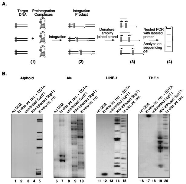
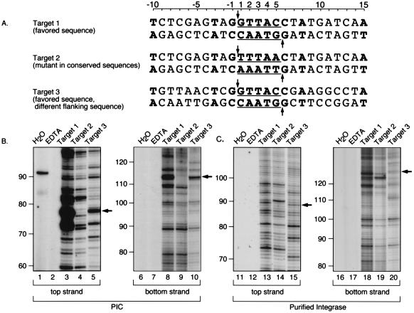
Integration of retroviral cDNA into host chromosomal DNA is an essential and distinctive step in viral replication. Despite considerable study, the host determinants of sites for integration have not been fully clarified. To investigate integration site selection in vivo, we used two approaches. (i) We have analyzed the host sequences flanking 61 human immunodeficiency virus type 1 (HIV-1) integration sites made by experimental infection and compared them to a library of 104 control sequences. (ii) We have also analyzed HIV-1 integration frequencies near several human repeated-sequence DNA families, using a repeat-specific PCR-based assay. At odds with previous reports from smaller-scale studies, we found no strong biases either for or against integration near repetitive sequences such as Alu or LINE-1 elements. We also did not find a clear bias for integration in transcription units as proposed previously, although transcription units were found somewhat more frequently near integration sites than near controls. However, we did find that centromeric alphoid repeats were selectively absent at integration sites. The repeat-specific PCR-based assay also indicated that alphoid repeats were disfavored for integration in vivo but not as naked DNA in vitro. Evidently the distinctive DNA organization at centromeres disfavors cDNA integration. We also found a weak consensus sequence for host DNA at integration sites, and assays of integration in vitro indicated that this sequence is favored as naked DNA, revealing in addition an influence of target primary sequence.


Figures

FIG. 1
FIG. 1
Cloning strategies for constructing integration site libraries. See the text for details and Table 1 for the sequences of oligonucleotides used.
FIG. 2
FIG. 2
Consensus sequence at the junctions between HIV cDNA and host DNA and the mechanism of generation of the host sequence duplication. (A) Integration pathway. HIV cDNA is shown as the curved line in part 1. Two nucleotides are removed from each 3′ end of the cDNA (part 2). Host target DNA is shown as a straight line. The host DNA that becomes duplicated is indicated by the numbers 1 to 5. The recessed 3′ ends of the cDNA are then attached to protruding 5′ ends in the target DNA (part 3), and the integration intermediate melts to yield single-stranded gaps at each end (part 4). The in vitro integration reactions with PICs stop at this stage. Repair of the DNA gaps at each host-virus DNA junction results in the production of the 5-bp duplication of target DNA (part 5). (B) Tabulation of the host sequence inferred to be duplicated in our integration site collection. HIV cDNA is joined to target DNA just 5′ of position 1, as illustrated, and similarly on the other strand. Sixty-six duplications are included in this compilation, 61 from the sites listed in Table 2 and 5 additional integration sites with the following duplication sequences: 5′-AGAGT-3′, 5′-GGTAC-3′, 5′-AACAT-3′, 5′-GTAAC-3′, 5′-AATGT-3′ (data not shown).
FIG. 3
FIG. 3
Analysis of integration sites near several repeat families using a PCR-based assay. (A) Diagram of the PCR method used to analyze integration sites. Primer binding sites are shown as gray rectangles. Part 1 illustrates either integration in vivo into cellular chromosomes or integration in vitro into deproteinized DNA. Products of integration reactions in vitro differ from products made in vivo in that only the former has the DNA breaks indicated in part 2 (the gapped integration intermediate is quickly repaired in vivo). In part 4, the three bands on the sequencing gel arose from three different integration events. (B) Results of PCR assays using primers complementary to alphoid repeats (lanes 1 to 5), Alu elements (lanes 6 to 10), LINE-1 elements (lanes 11 to 14), and THE 1 elements (lanes 16 to 20). The presence of a ladder of bands indicates that the template DNA contained HIV cDNA integrated near the repeat family specified. Lanes: 1, 6, 11, and 16, control amplification reactions with no added template; 2, 7, 12, and 17, amplification of inactive PICs and SupT1 DNA; 3, 8, 13, and 18, amplification from uninfected SupT1 DNA; 4, 9, 14, and 19, amplification of DNA from HIV-1 infected SupT1 cells; 5, 10, 15, and 20, amplification of deproteinized DNA that had been incubated with active PICs in vitro. Cellular DNA was detectable as a contaminant of the PIC preparations (data not shown); cellular DNA might have served as an integration target during PIC preparation or participated in recombination during PCR, possibly giving rise to the artifactual bands in lanes 7 and 12.
FIG. 4
FIG. 4
A conserved sequence at integration sites and analysis of integration at such sites in vitro. (A) Integration target sites tested. The host sequences duplicated upon integration are underlined; the points at which covalent strand transfer takes place on each strand are indicated by arrows; bases favored at integration sites are in boldface type. (B) Integration into targets 1 to 3 directed by PICs. Lanes: 1 and 6, H2O instead of template; 2 and 7, EDTA added to integration reactions. 3 and 8, target 1; 4 and 9, target 2; 5 and 10, target 3. Arrows indicate the location of the expected integration hotspots (5′ of position 1 on the top strand and 5′ of position 5 on the bottom strand). (C) Integration into targets 1 to 3 directed by purified HIV-1 integrase. Lanes 11 to 20 correspond to lanes 1 to 10, respectively, in panel B. Sizes were assigned by coelectrophoresis adjacent to several DNA sequencing ladders generated by the Sanger method.

Similar articles

Cited by

References

    1. Altschul S F, Madden T L, Schaffer A A, Zhang J, Zhang Z, Miller W, Lipman D J. Gapped BLAST and PSI-BLAST: a new generation of protein database search programs. Nucleic Acids Res. 1997;25:3389–3402. - PMC - PubMed
    1. Boeke J D. Transposable elements in Saccharomyces cerevisiae. In: Berg D E, Howe M M, editors. Mobile DNA. Washington, D.C: American Society for Microbiology; 1989. pp. 335–374.
    1. Bor Y-C, Bushman F, Orgel L. In vitro integration of human immunodeficiency virus type 1 cDNA into targets containing protein-induced bends. Proc Natl Acad Sci USA. 1995;92:10334–10338. - PMC - PubMed
    1. Bor Y-C, Miller M, Bushman F, Orgel L. Target sequence preferences of HIV-1 integration complexes in vitro. Virology. 1996;222:238–242. - PubMed
    1. Brown P O, Bowerman B, Varmus H E, Bishop J M. Correct integration of retroviral DNA in vitro. Cell. 1987;49:347–356. - PubMed

Publication types

LinkOut - more resources